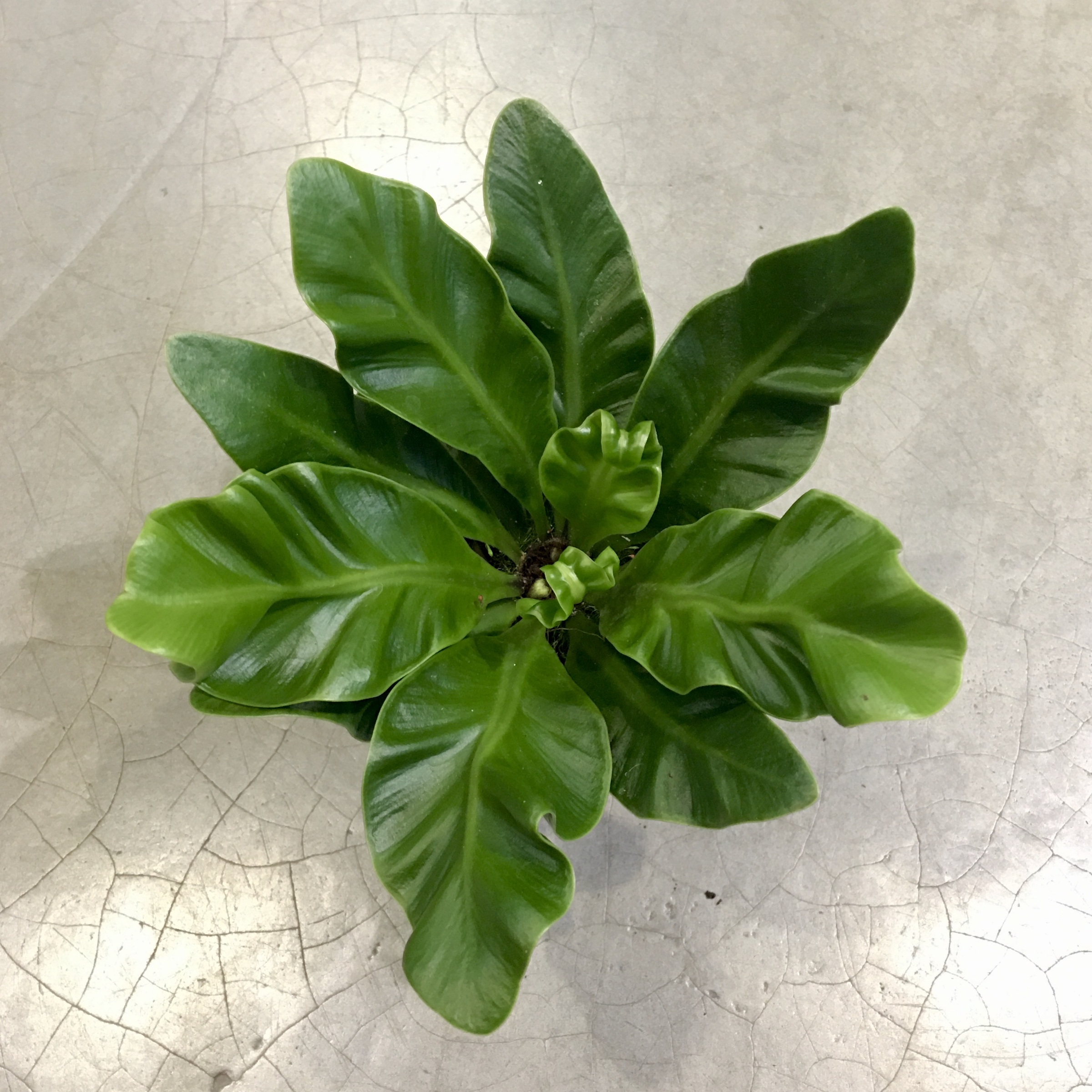
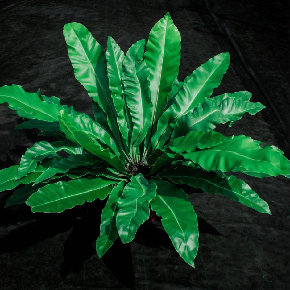
ASPLENIUM NIDUS Bird's Nest Fern 'Crispy Wave' Tulbagh Tree & Plant Nursery
Especially in the kidney-shaped stomata of the fern Asplenium nidus, callose actively participates in the mechanism of opening and closure of the stomatal pore. Scope The present review briefly presents and discusses recent findings concerning the distribution and role of callose in the kidney-shaped stomata of the dicotyledon Vigna sinensis as.
Asplenium nidus Botanically
In stomata of the fern Asplenium nidus, callose is implicated in stomatal pore formation, guard cell wall thickenings formation as well as in stomatal function [28][29] [30]. The deposition of.
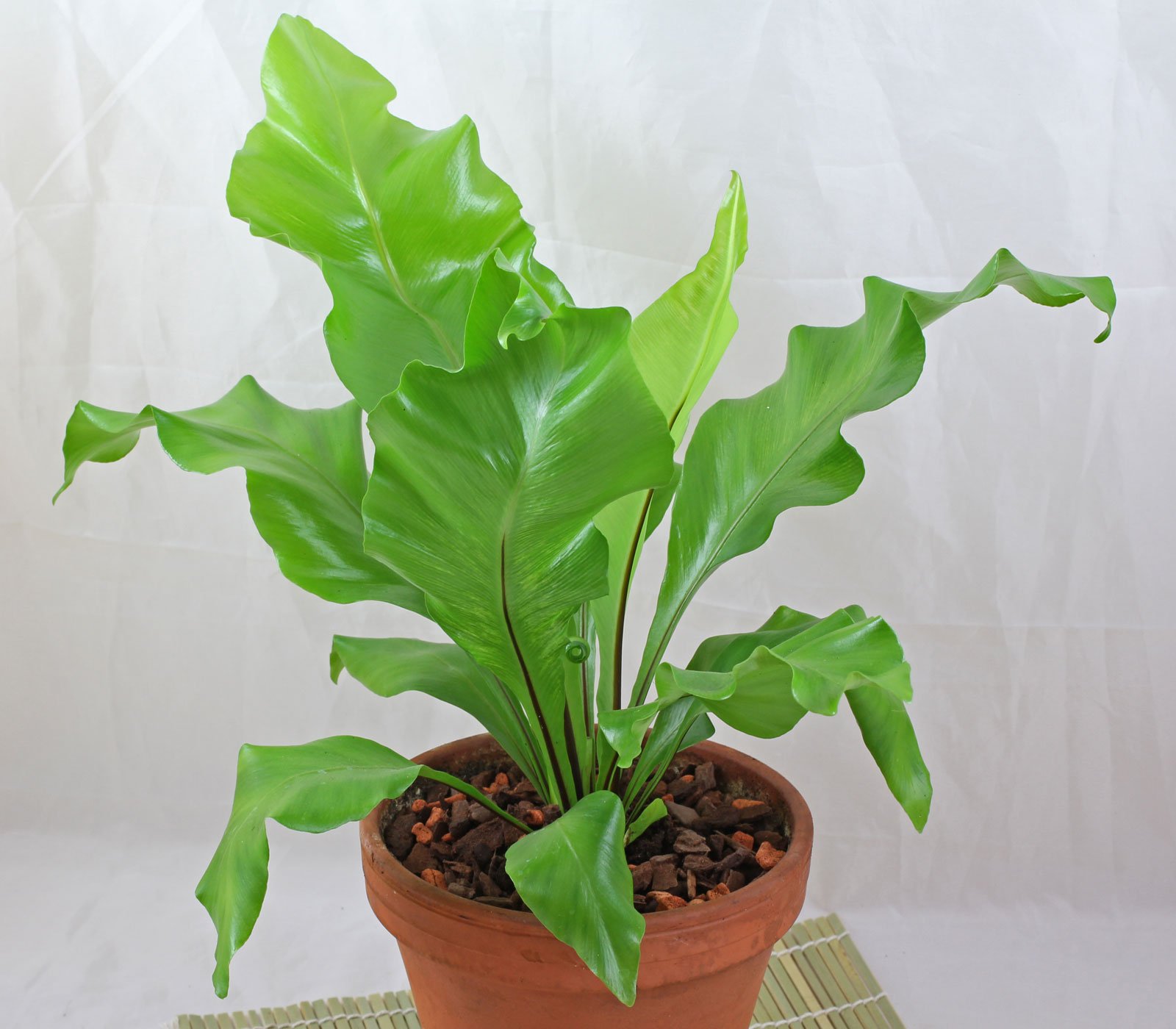
Asplenium nidus
Stomatal-pore formation in the fern Asplenium nidus L. commences in postcytokinetic guard cells at the mid-region of the ventral wall, before the deposition of any cellulosic wall material on it, by the local movement of the adjacent plasmalemmata apart from each other. In this way a rudimentary "internal stomatal pore" is formed. At this stage the ventral wall exhibits an undulated.

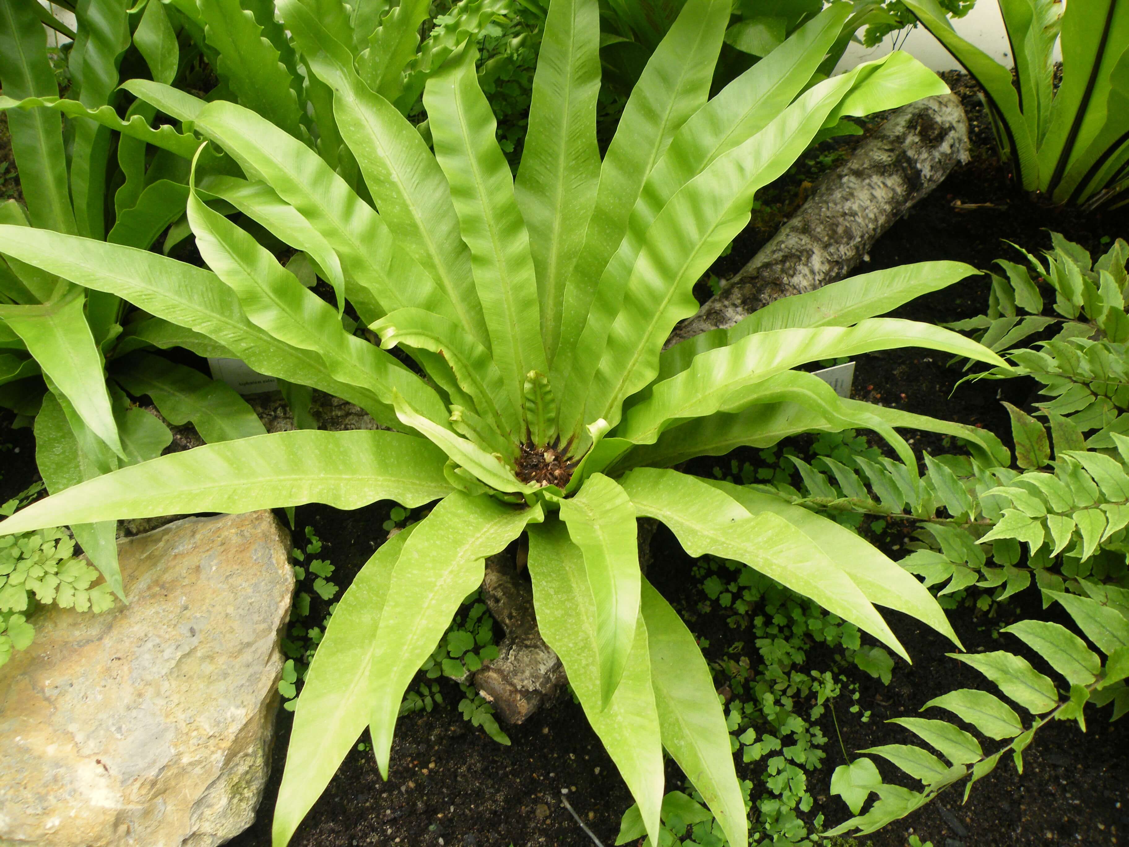
Asplenium nidus L. Plants of the World Online Kew Science
The six species of vascular plants chosen for this study cover a broad structural, ecophysiological and evolutionary spectrum: ferns (Asplenium nidus and Platycerium bifurcatum) and angiosperms (Arabidopsis thaliana and Commelina erecta) with kidney-shaped stomata, and grasses (angiosperms, family Poaceae) with dumbbell-shaped stomata (Sorghum.
Asplenium nidus Ferns and Lycophytes of the World
Although the cellulose microfibril organization in guard cell (GC) walls play a crucial role in the mechanism of the stomatal function, recent work showed that matrix cell wall materials are also involved. Especially in the kidney-shaped stomata of the fern Asplenium nidus, callose actively participates in the mechanism of opening and closure.
September 2017 Page 17 PLANT STOMATA ENCYCLOPEDIA
Callose implication in stomatal opening and closure in the fern Asplenium nidus

Asplenium nidus (6cm Pot) grow urban.
Background Although the cellulose microfibril organization in guard cell (GC) walls play a crucial role in the mechanism of the stomatal function, recent work showed that matrix cell wall materials are also involved. Especially in the kidney-shaped stomata of the fern Asplenium nidus, callose actively participates in the mechanism of opening and closure of the stomatal pore. Scope The present.

Asplenium nidus L. a photo on Flickriver
Background and aims: The pattern of callose deposition was followed in developing stomata of the fern Asplenium nidus to investigate the role of this polysaccharide in guard cell (GC) wall differentiation and stomatal pore formation. Methods: Callose was localized by aniline blue staining and immunolabelling using an antibody against (1 --> 3)-beta-d-glucan.
Asplenium nidus L. Plants of the World Online Kew Science
Microtubule and actin filament organization during stomatal morphogenesis in the fern Asplenium nidus. II. Guard cells - Volume 141 Issue 2. Skip to main content Accessibility help We use cookies to distinguish you from other users and to provide you with a better experience on our websites.
FileAspleniumnidus.JPG Wikipedia
ing in the fern Asplenium nidus was investigated by examination of the pattern of callose deposition in open and closed stomata, and by examination of the effects of callose degradation and inhibition or induction of callose synthesis in stomatal movement. • Callose was identified with aniline blue staining and a callose antibody and

Asplenium nidus Bessey Greenhouse (Richard W. Pohl Conservatory)
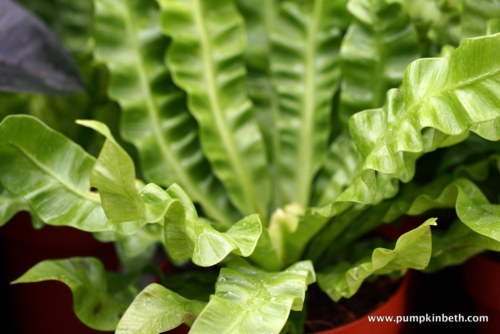
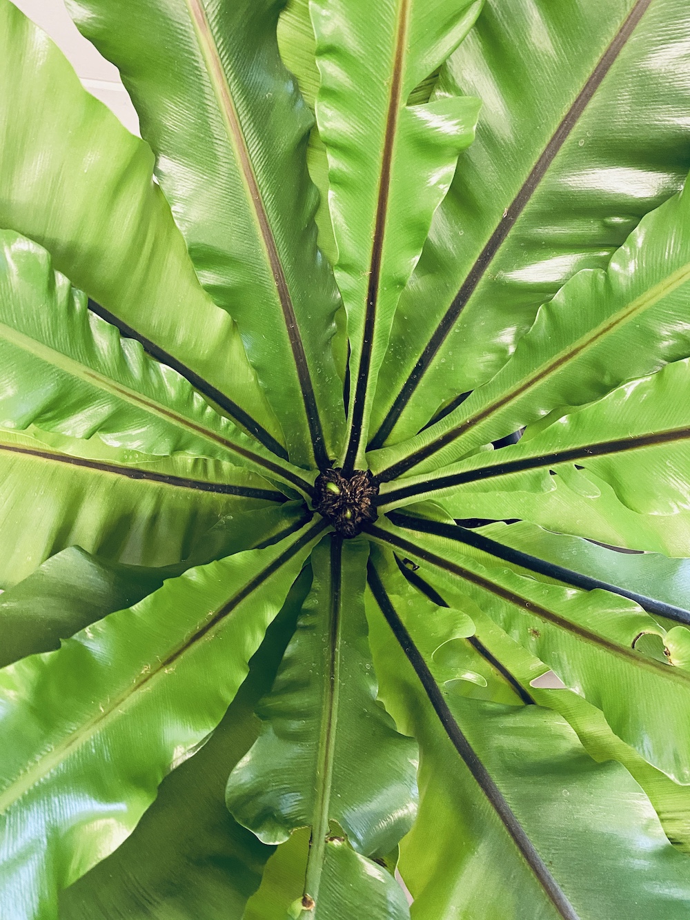
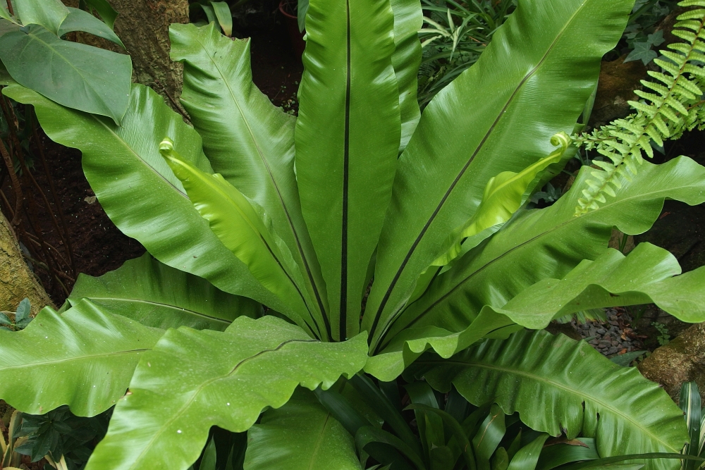
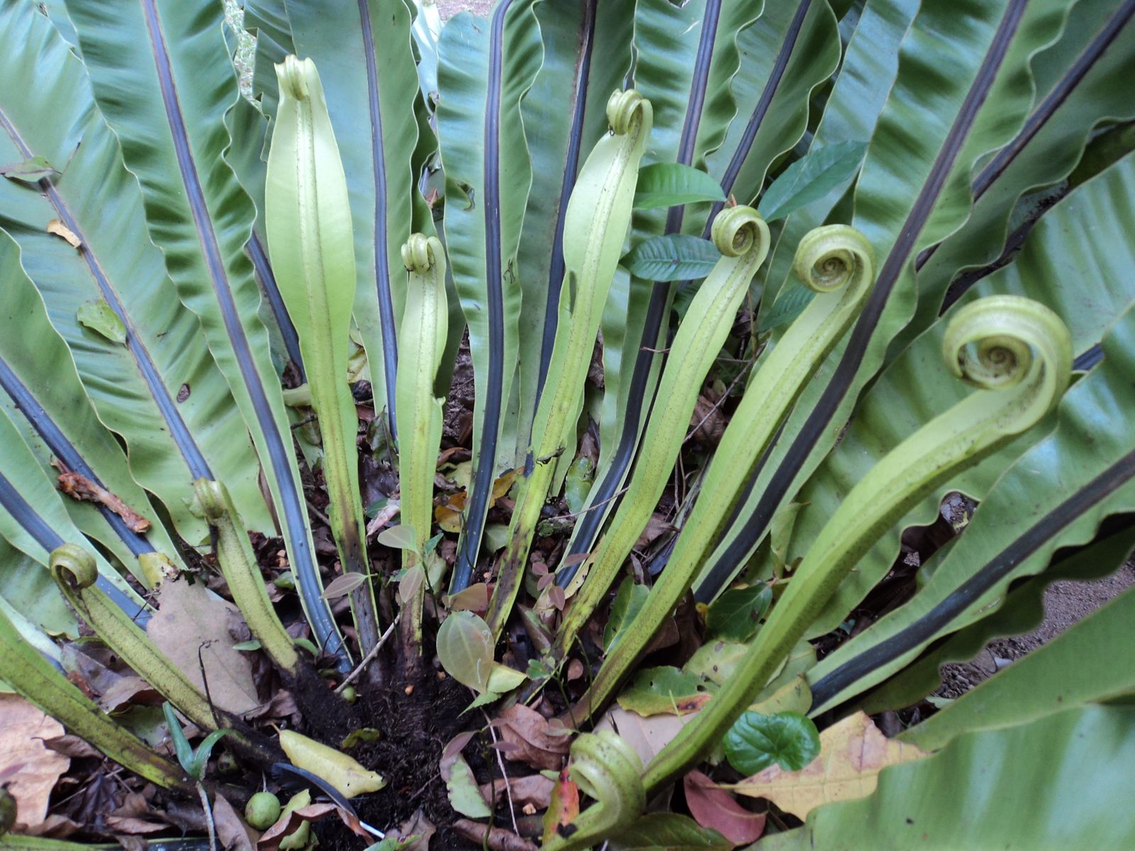
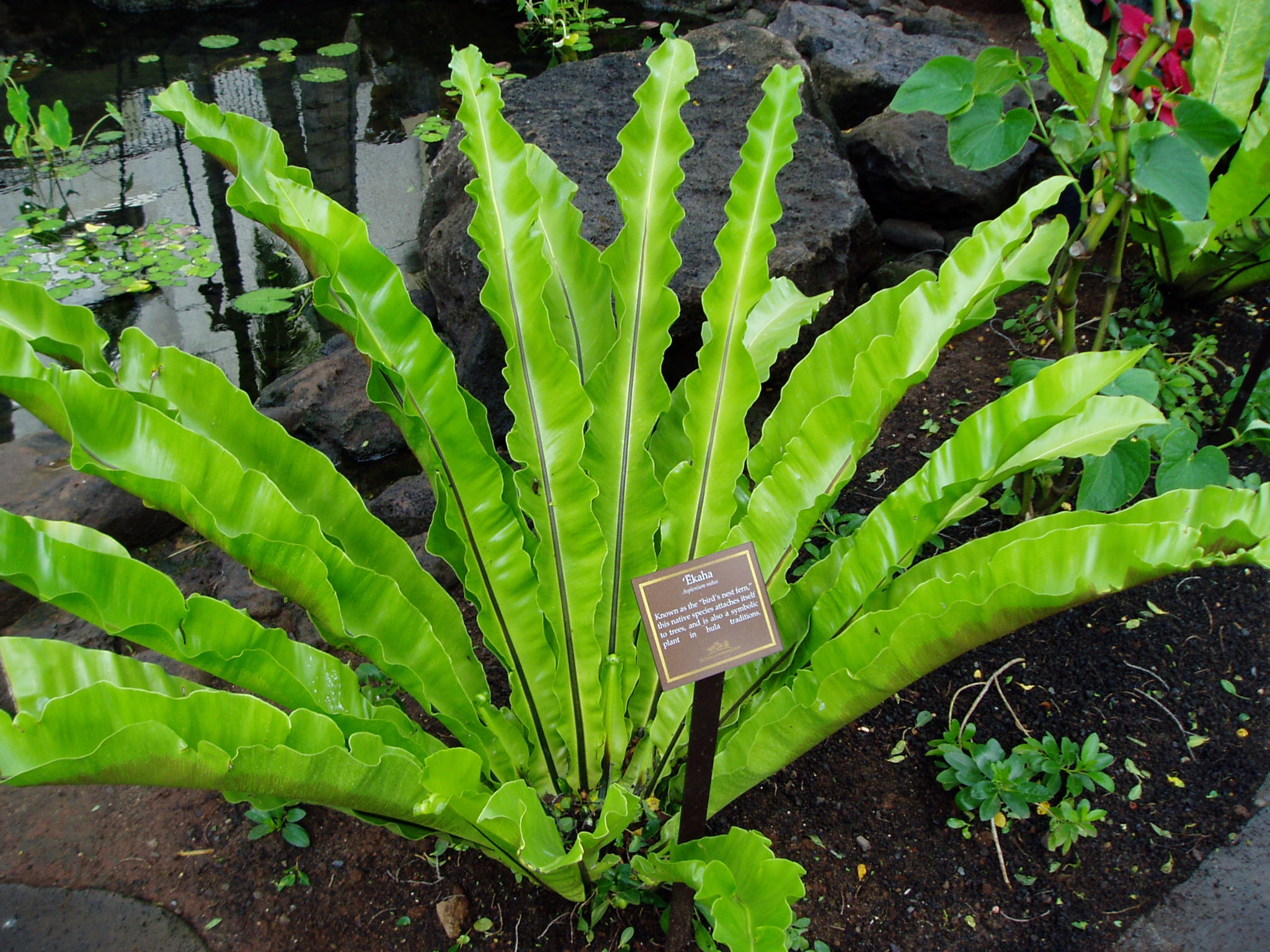
Asplenium nidus is an epiphytic fern with large simple leaves. Because A. nidus lacks the good taxonomic characters available for species recognition, multiple cryptic species may exist within A.
Asplenium nidus Finca Drácula
The newly-formed guard cell mother cells (GMCs) ofAsplenium nidus are small, lens-shaped and are formed by one or two asymmetrical divisions. Their growth axis is parallel to the plane of their future division, a process during which the internal periclinal wall (IPW) is detached from the partner wall of the underlying cell(s). This oriented GMC expansion occurs transversely to a microfibril.
Asplenium nidus Riverside Garden Centre
Callose in living Asplenium nidus L. stomata was localized using aniline blue staining (O'Brien and McCully, 1981). Callose was also labelled in fixed free-hand and semi-thin sections using a monoclonal antibody against (1 → 3)-β-d-glucans (Meikle et al., 1991; Ferguson et al., 1998). Treatments

Asplenium nidus L. Plants of the World Online Kew Science
The involvement of callose in the mechanism of stomatal pore opening and closing in the fern Asplenium nidus was investigated by examination of the pattern of callose deposition in open and closed stomata, and by examination of the effects of callose degradation and inhibition or induction of callose synthesis in stomatal movement.. Callose was identified with aniline blue staining and a.
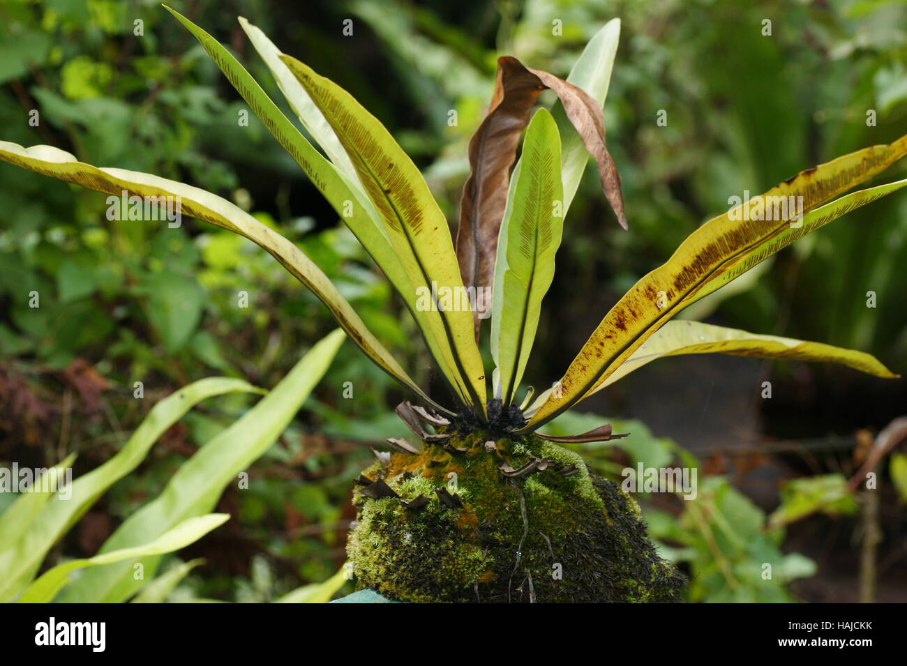
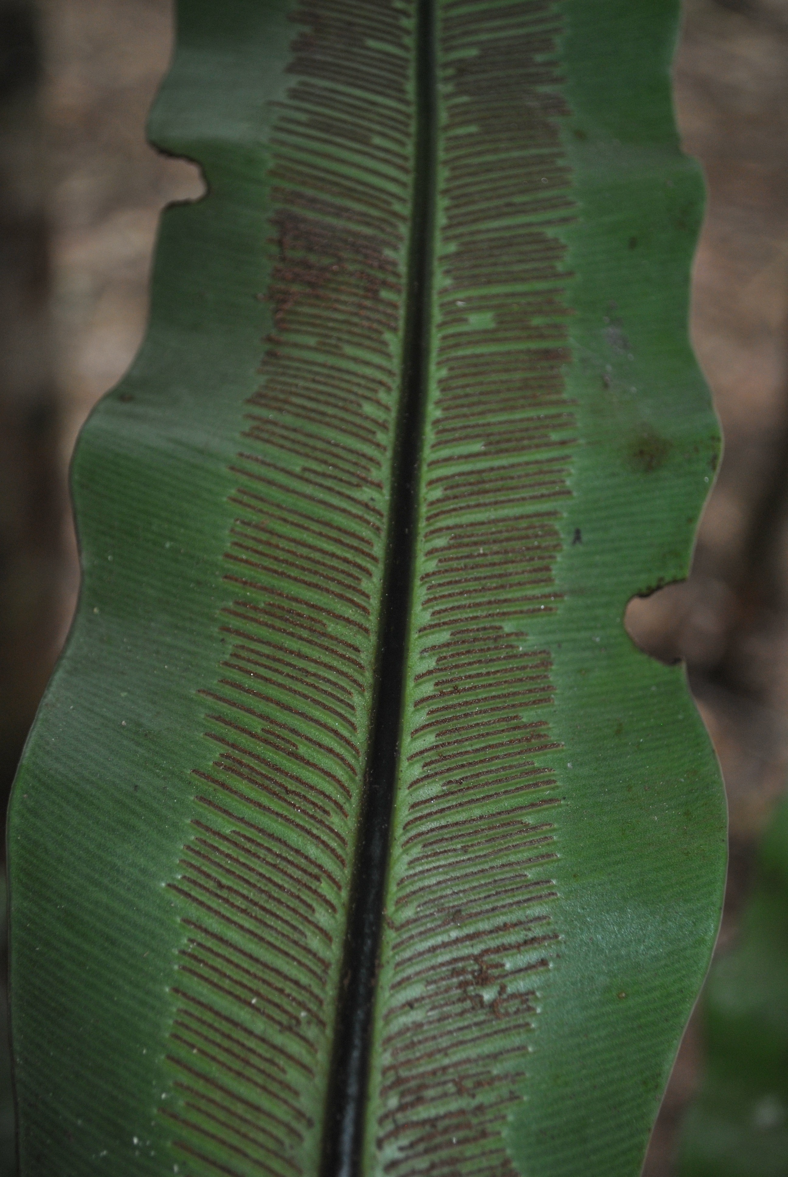
Asplenium nidus, bird'snest fern, nest fern. Spores develop in sori on the underside of the
Comparative morphological and foliar anatomical studies were carried out on three species of Asplenium: Asplenium nidus L., Asplenium scolopendrium (L.) Newn and Asplenium barterii Hook with a view to identifying important morphological and anatomical characters that can be employed in the separation of the species. Mature healthy plants of each species were collected from Erin Ijesa Waterfall.