
Normal Sagittal and Coronal Suture Widths by Using CT Imaging American Journal of Neuroradiology
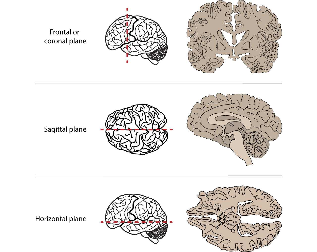
The sagittal suture extends posteriorly from the coronal suture, running along the midline at the top of the skull in the sagittal plane of section. It unites the right and left parietal bones. On the posterior skull, the sagittal suture terminates by joining the lambdoid suture. The lambdoid suture extends downward and laterally to either side.
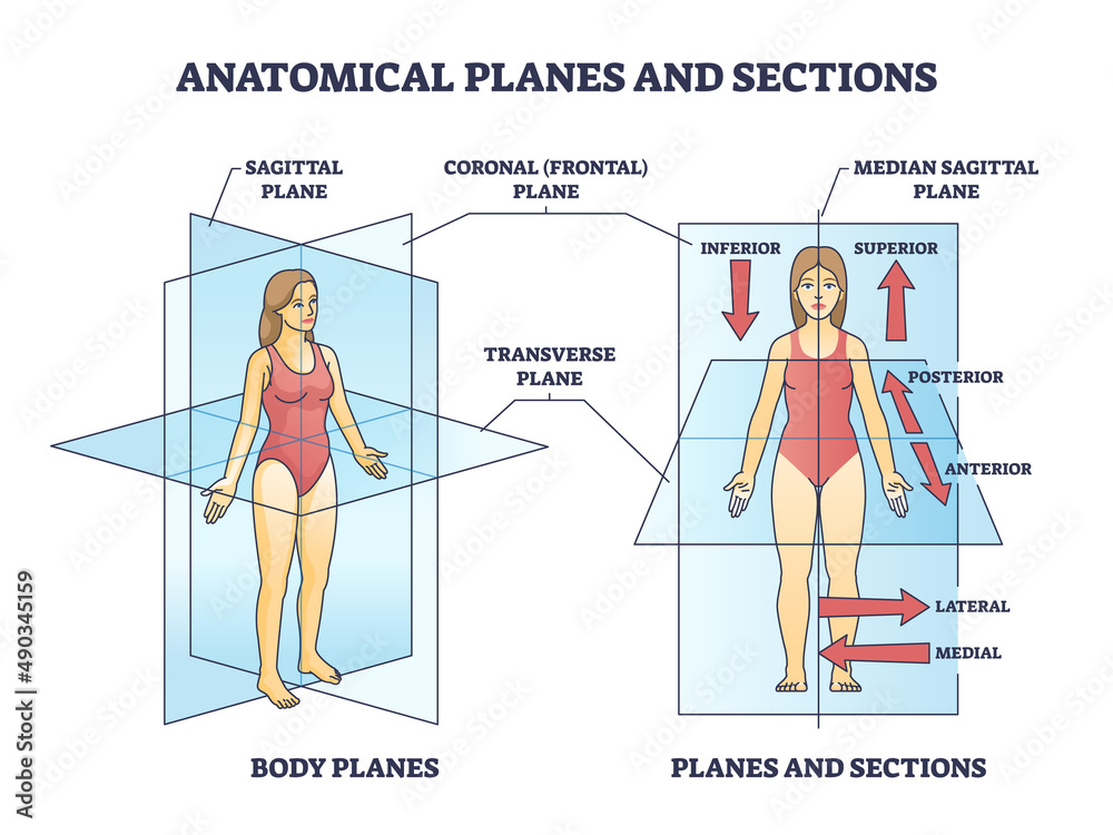
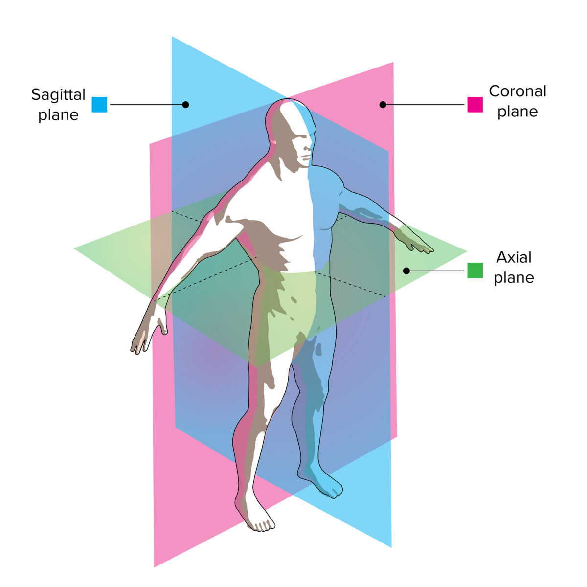
Anatomical planes or sections for human medical body division outline diagram. Labeled
The coronal suture runs from side to side across the skull, within the coronal plane of section (see Figure 7.3.3). It joins the frontal bone to the right and left parietal bones. The sagittal suture extends posteriorly from the coronal suture at the intersection called bregma, running along the midline at the top of the skull in the sagittal.

Axial Skeleton Learn Skeleton Anatomy
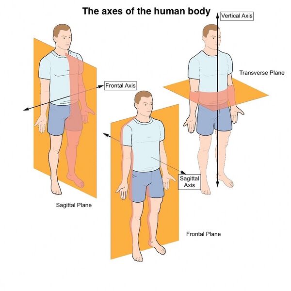
Sagittal and coronal planes are also referred to as longitudinal planes as they make a right angle to the transverse plane. Although motion is a combination of different movements, it can be classified depending upon the anatomical plane where it has occurred. To describe these planes, consider a person standing with an upright stance.
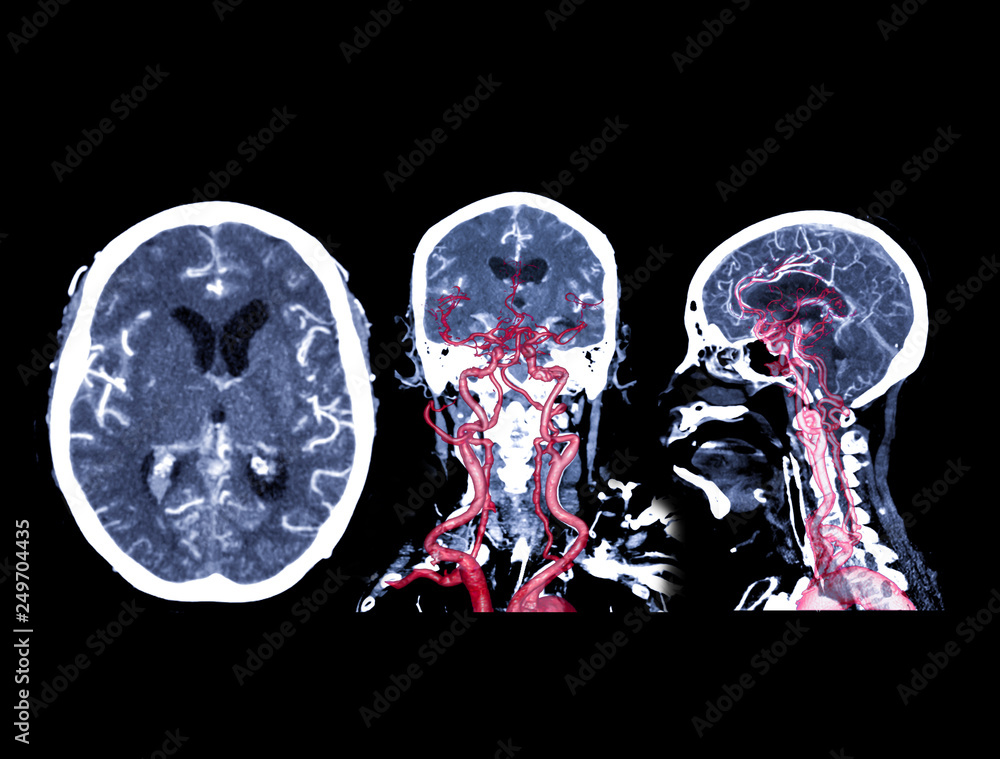
Collection of CT angiography of the brain or CTA brain comparison Axial , Coronal and Sagittal
Figure 5.1.1 5.1. 1: The axial skeleton highlighted in blue (Public Domain, LadyofHats Mariana Ruiz Villarreal, Wikimedia) The axial skeleton consists of the bones of the skull, the bones of the inner ear (known as ossicles), the hyoid bone in the throat, and the bones of the vertebral column, including the sacrum and coccyx bones in the center.

Normal Sagittal and Coronal Suture Widths by Using CT Imaging American Journal of Neuroradiology
Anatomical position for a human is when the human stands up, faces forward, has arms extended, and has palms facing out. Figure 1.2.1 1.2. 1 : These two people are both in anatomical position. (CC-BY, Open Stax ) When referencing a structure that is on one side of the body or the other, we use the terms "anatomical right" and "anatomical.

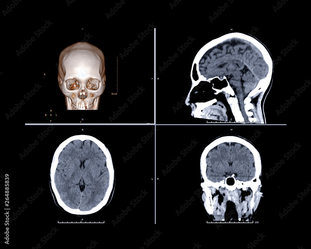
Imaging of the Head and Brain Concise Medical Knowledge
UCSF Radiologist Dr. Dillon describes how radiologists read images. The different planes that Radiologists use are axial (divides the body into top and bottom halves), coronal (perpendicular), and sagittal (midline of the body). Radiologists call images that are axial or coronal view differently as they reverse left and right. Radiologists view CT and MR as if they are looking from the feet.
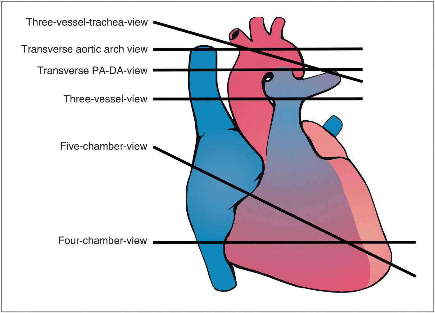
The Great Vessels Axial, Oblique, and Sagittal Views Obgyn Key
A coronal or frontal plane divides the body into dorsal and ventral (back and front, or posterior and anterior) portions. A transverse plane, also known as an axial plane or cross-section, divides the body into cranial and caudal (head and tail) portions. A sagittal plane divides the body into sinister and dexter (left and right) portions.
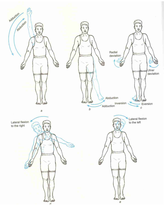
Cardinal Planes and Axes of Movement Physiopedia
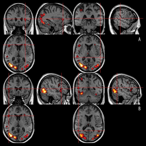
However, intracerebral reference points for the axial, sagittal, and coronal planes of brain have not been standardized in anatomical sections or radiological images. We made 2,343 serially-sectioned images of a cadaver head with 0.1 mm intervals, 0.1 mm pixel size, and 48 bit color and obtained axial, sagittal, and coronal images based on the.
Cardinal Planes and Axes of Movement Physiopedia
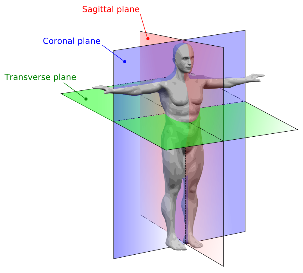
An anatomical plane is a hypothetical plane used to transect the body, in order to describe the location of structures or the direction of movements. In human and non-human anatomy, three principal planes are used: The sagittal plane or lateral plane (longitudinal, anteroposterior) is a plane parallel to the sagittal suture.It divides the body into left and right.
Sagittal, coronal and axial views of the Broca’s and Openi
CT evaluation of diffuse infiltrative lung disease: dose considerations and optimal technique. J Thorac Imaging. 2009;24:252-259. HRCT Primer. Image Reconstruction Planes. Review the different image reconstruction planes, which include axial, coronal, and sagittal planes and are made possible using volumetric acquisition CT.
T1weighted sagittal, axial, and coronal images as exam Openi
The coronal or frontal planes divide the body into front and back (also called dorsal and ventral or posterior and anterior) sections and are x-y planes. The transvers planes, also known as the.
Anatomical Location Anatomy and Physiology I
A coronal sequence should be perpendicular to the short axis of the glenoid articular surface (Figure 12-1A), while the sagittal is parallel to the short axis of the glenoid articular surface (Figure 12-1B), and the axial is perpendicular to the long axis of the glenoid articular surface (Figure 12-1C). Both fat- and fluid-sensitive sequences.
Imagenología de la Cabeza y el Cerebro Concise Medical Knowledge
Consider which poses you tend to practice at home and which ones you avoid. STEP 2 Determine the primary plane for each of the poses on your lists. STEP 3 Name the planes in which you seem to be most and least comfortable. STEP 4 Create a list of poses from your least favorite plane, and plan to practice these poses several times a week.
Anatomical Terminology Foundations of Neuroscience
High field strength 1.5T or 3T scanners and dedicated extremity coils provide adequate signal-to-noise ratio and the spatial resolution required for proper imaging of the complex regional anatomy of the ankle and foot. Standard MRI planes for ankle and foot include sagittal, coronal, and axial planes. For examination of the forefoot, imaging.
CT brain trauma comparison 3D rendering image , sagittal , axial and coronal view for screening
The plane of the angle of the sagittal image is parallel to the interhemispheric fissure. The angle of the coronal image is usually parallel to the bed of the MR equipment or parallel to the brainstem. However, there are six angles for the axial image. This is because axial imaging has a long history since the CT era.

Identification of anatomical planes applied to brain Diagram Quizlet
Coronal (frontal) plane: separates the front (anterior) and back ( posterior) of the body. Sagittal (longitudinal) plane: separates the left and right sides of the body. Transverse (axial) plane.
