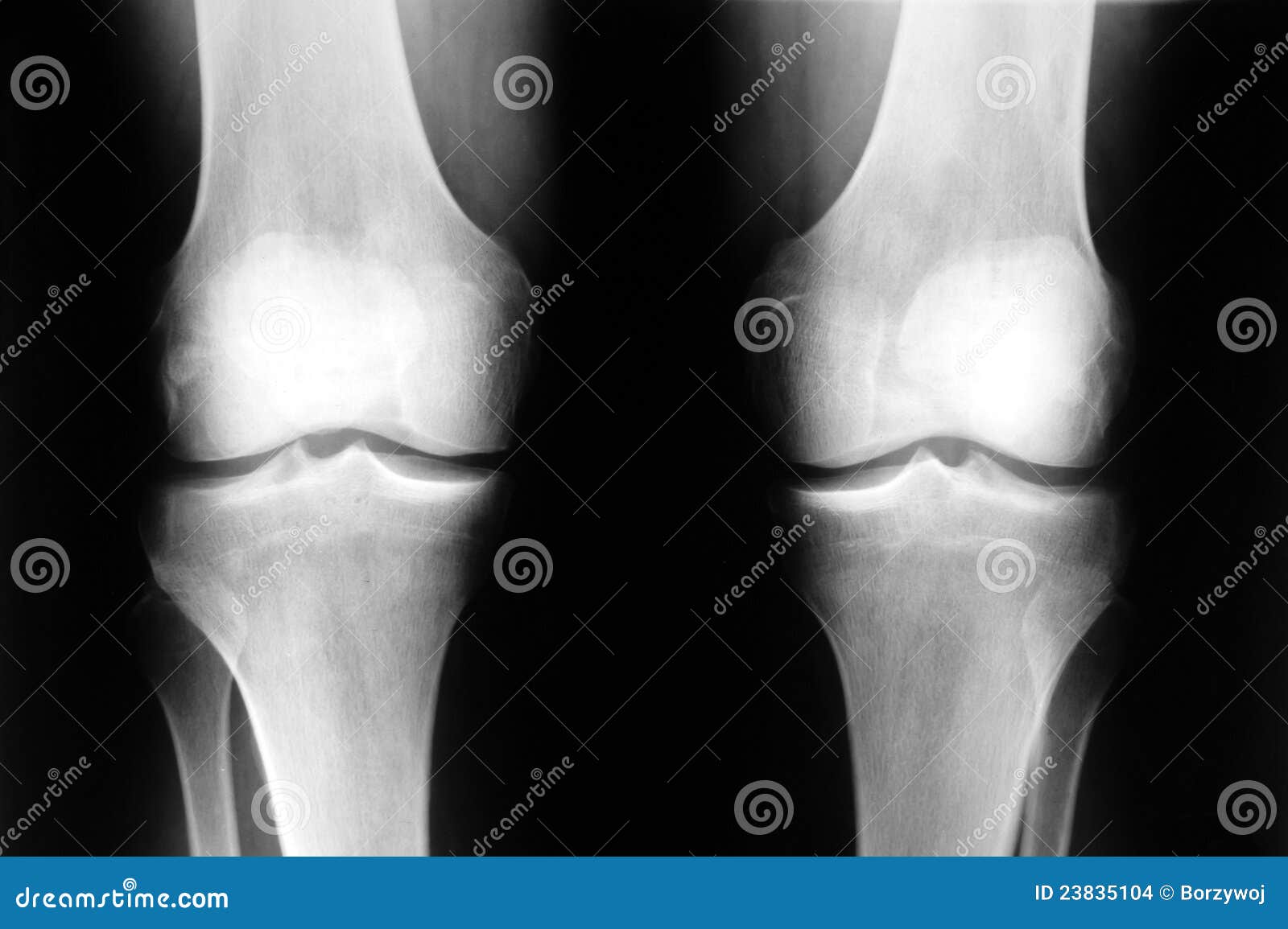
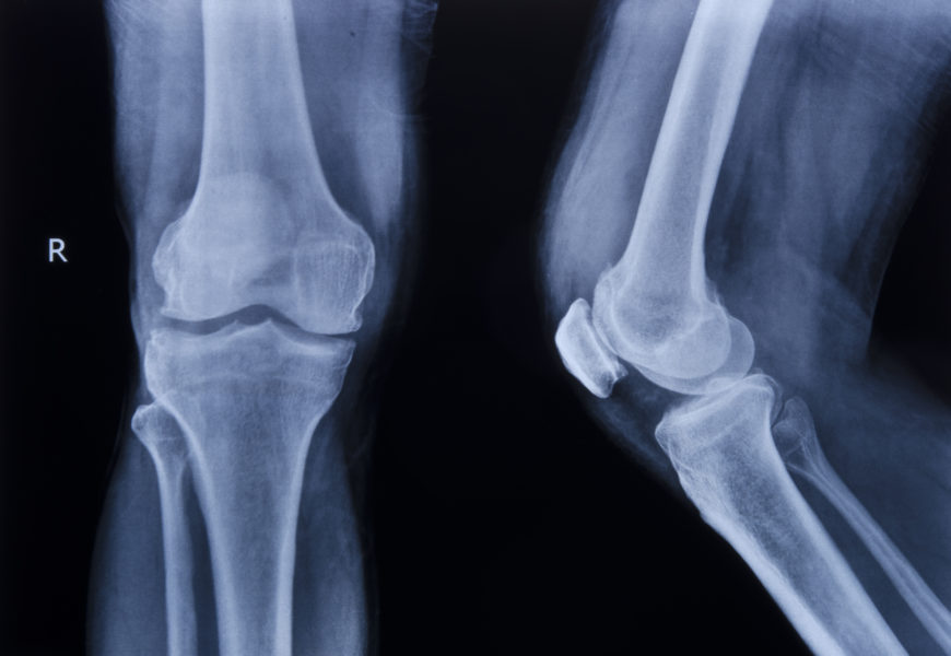
Knee stock photo. Image of rontgen, real, human, sick 23835104
Prosedur pemeriksaan rontgen dibagi menjadi tiga tahapan yakni sebelum foto, selama foto, dan sesudah foto. Ini ulasan untuk masing-masing tahapan: 1. Sebelum Foto. Kamu dianjurkan untuk berpuasa atau berhenti menggunakan obat-obatan. Kamu juga harus melepas perhiasan atau aksesoris yang terbuat dari logam.

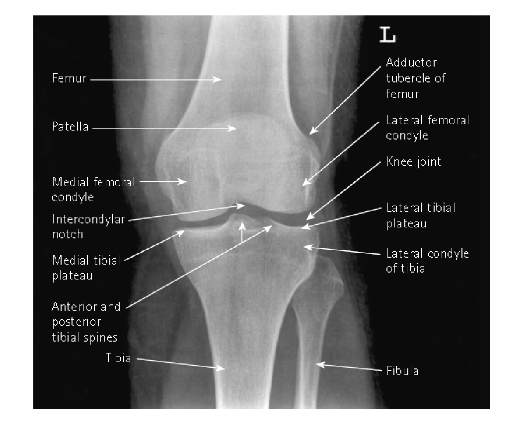
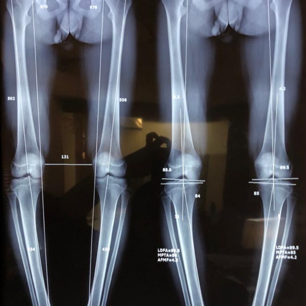
Radiographic Anatomy Knee AP Radiology student, Medical knowledge, Medical anatomy
2012 ICD-9-CM Procedure Code 88.02. Other Abdomen Tomography. 88.02 is a specific code and is valid to identify a procedure. 2012 ICD-9-CM Procedure Code 88.03. Sinogram Of Abdominal Wall. 88.03 is a specific code and is valid to identify a procedure. 2012 ICD-9-CM Procedure Code 88.04. Abdominal Lymphangiogram.
.png)
Diagnostics Knee and Ankle Xrays — Taming the SRU
Citation, DOI, disclosures and article data. Blount disease refers to a local disturbance of growth of the medial aspect of the proximal tibial metaphysis and/or epiphysis that results in tibia vara . The condition is commonly bilateral. Somewhat confusingly, "tibia vara" has been used in the literature as a synonym for Blount disease.

Rechtes Knie Röntgen Stockfotografie Alamy
Indications. The Rosenberg view is performed for any patient with a suspicion of knee osteoarthritis. It consists of a PA radiograph with weight-bearing and 45° of knee flexion. It is more sensitive than standard weight-bearing radiographs for the detection of joint space narrowing 1.
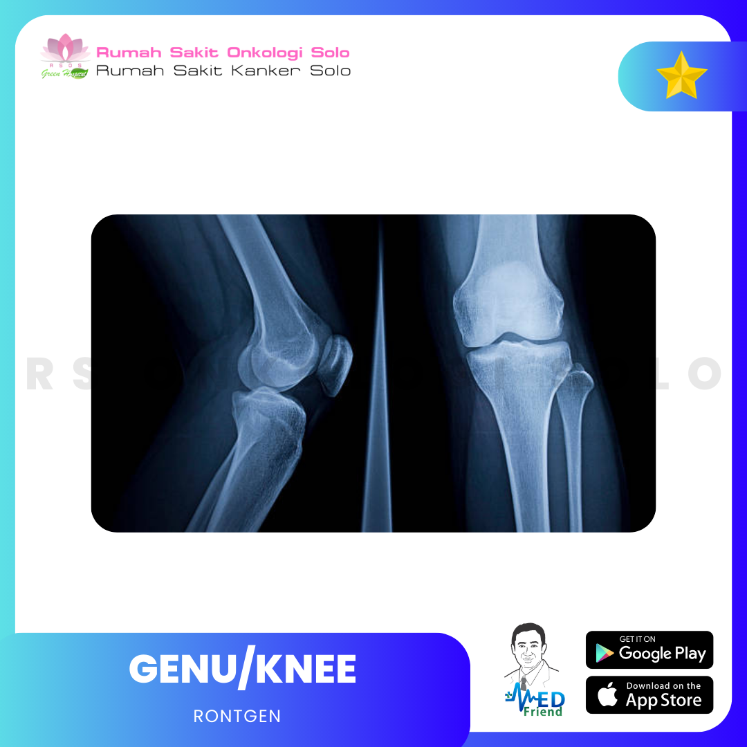
RONTGEN GENU/KNEE
2012 ICD-9-CM Procedure Code 87.1. Other X-Ray Of Face, Head, And Neck. A child code below 87.1 with greater detail should be used. 2012 ICD-9-CM Procedure Code 87.11. Full-Mouth X-Ray Of Teeth. 87.11 is a specific code and is valid to identify a procedure. 2012 ICD-9-CM Procedure Code 87.12. Other Dental X-Ray.
Alami Osteoarthritis Genu Sinistra Usai Kecelakaan Halaman 1
the patient is semi-recumbent on the table holding a detector superior of the patella in the landscape orientation. patient's feet should be very close to the tube side of the bed (see technical factors) the knee is bent close to 30°. often a pillow or cushion should be placed behind the patient to assist them in maintaining this position.

TEKNIK PEMERIKSAAN KNEE JOINT
Serta sebelum pemeriksaan Anda akan diminta untuk meminum banyak air atau dan menahan kencing agar dapat terlihat gambaran yang bagus pada buli-buli (kandung kemih). Pemeriksaan dada proyeksi posterior anterior (PA) dilakukan dengan posisi berdiri, baju harus diturunkan sampai ke pinggang. Anda akan diminta untuk menahan nafas saat foto diambil.
TEKNIK PEMERIKSAAN KNEE JOINT
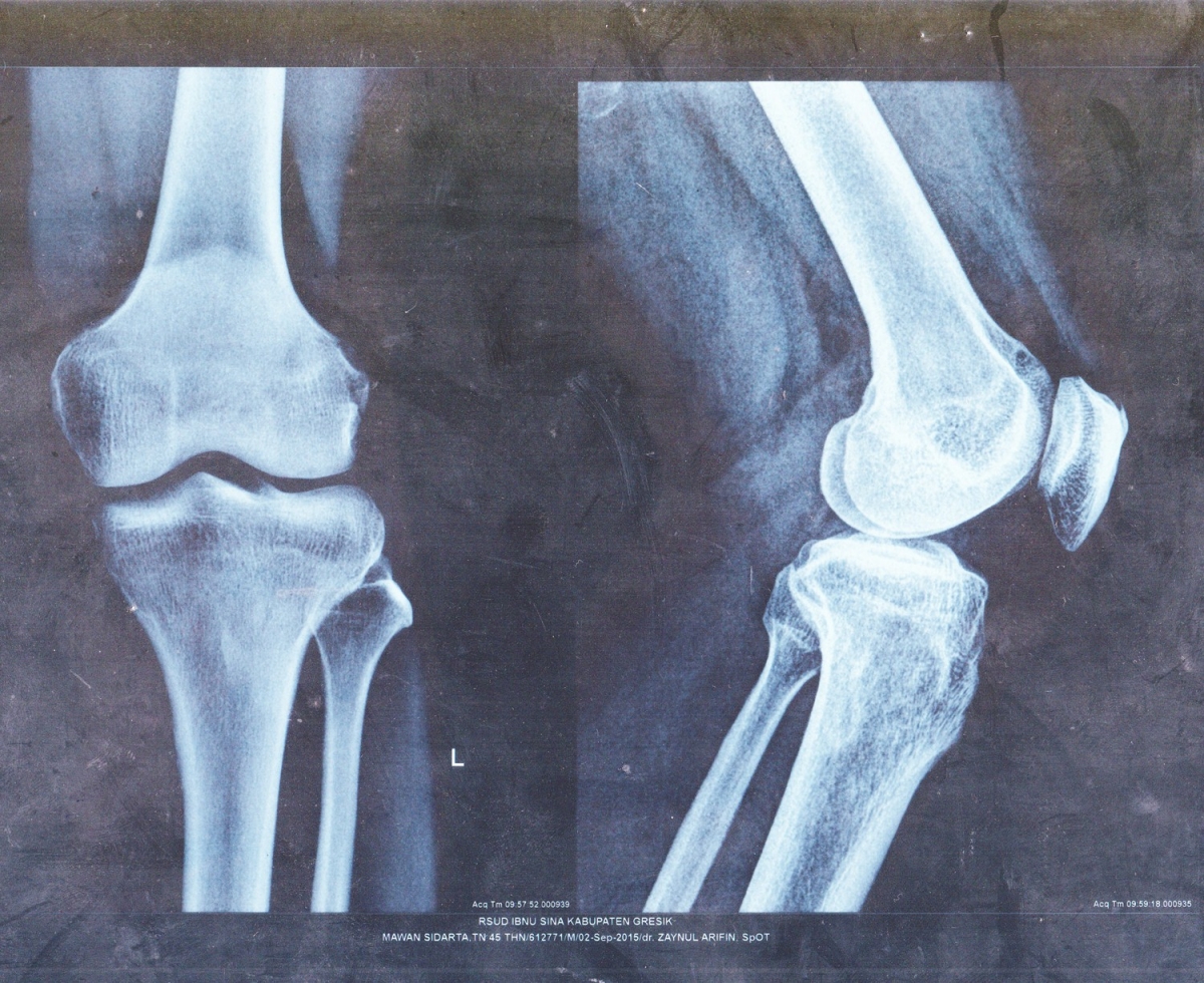
20 foto rontgen genu mengalami Post-TKR, satu foto mengalami Post-THR, dan 10 yang tidak terdapat foto rontgen dan intepretasi foto. Jadi, total sampel pada penelitian ini adalah 91 (74,6%) sampel foto rontgen regio genu. Hasil foto rontgen regio genu pada pasien Osteoartritis Primer di RSUP Sanglah

Asymptomatic Hyperuricemia Predictive of Radiographic Knee Osteoarthritis Rheumatology Advisor
Wilhelm Conrad Röntgen (born March 27, 1845, Lennep, Prussia [now Remscheid, Germany]—died February 10, 1923, Munich, Germany) physicist who received the first Nobel Prize for Physics, in 1901, for his discovery of X-rays, which heralded the age of modern physics and revolutionized diagnostic medicine. Röntgen studied at the Polytechnic in.
Genu Varum Farhad Farid
Malalignment of the vertebrae, in patients suspected of blunt spinal trauma, is the quintessential sign of spinal injury. Malalignment is obvious in displaced fractures and dislocations but is rarely considered in the diagnosis because of the obvious injury. In patients with more subtle injuries such as those limited to ligamentous structures.
How To Read Knee Mri Knee Mri Meniscus Tear Mri Images and Photos finder
Rontgen genu kanan: Rp 267.000: Rontgen genu kiri: Rp 267.000: Rontgen pedis kanan: Rp 306.000: Rontgen pedis kiri: Rp 306.000: Rontgen telapak kaki: Rp 200.000: Biaya di atas bisa berubah sewaktu-waktu tergantung kebijakan Klinik Laboratorium Prodia. Kesimpulan.
Knee Radiology Made Surprisingly Simple Digital Teaching
Setidaknya 2 dari 3 kriteria berikut: LED < 20 mm/jam, osteofit femoral atau asetabular, penyempitan celah sendi (superior, aksial, atau medial) [16] Diagnosis Banding. Diagnosis lain yang harus dipikirkan dan disingkirkan pada kasus yang dicurigai osteoarthritis adalah gout dan rheumatoid arthritis. Gout.

KUMPULAN SOP (STANDAR OPERASIONAL PROSEDUR) SOP Radiologi Cara & Tekhnik Foto / Pemeriksaan
Wilhelm Conrad Röntgen (/ ˈ r ɛ n t ɡ ə n,-dʒ ə n, ˈ r ʌ n t-/; German pronunciation: [ˈvɪlhɛlm ˈʁœntɡən] ⓘ; 27 March 1845 - 10 February 1923) was a German mechanical engineer and physicist, who, on 8 November 1895, produced and detected electromagnetic radiation in a wavelength range known as X-rays or Röntgen rays, an achievement that earned him the inaugural Nobel.
Röntgen der beiden menschlichen Knie (normale Knie Stockfotografie Alamy
88 Other Diagnostic Radiology And Related Techniques. 88.0 Soft Tissue X-Ray Of Abdomen. 88.01 Computerized axial tomography of abdomen convert 88.01 to ICD-10-PCS. 88.02 Other abdomen tomography convert 88.02 to ICD-10-PCS. 88.03 Sinogram of abdominal wall convert 88.03 to ICD-10-PCS.

Victory Sports Medicine & Orthopedics Knee XRay Victory Sports Medicine & Orthopedics
The hallmarks of knee osteoarthritis are the same for most other joints 6: joint space narrowing. usually asymmetric, typically of the medial tibiofemoral compartment, and/or patellofemoral compartment 3. <3 mm on weight-bearing knee radiographs is considered a finding of absolute joint space narrowing with a normal joint space >5 mm 7.

Roentgen Unit Stock Photos, Pictures & RoyaltyFree Images iStock
Pemeriksaan rontgen genu (lutut) tersebut dilakukan untuk mengetahui adanya keluhan pada lutut misalnya adakah gangguan pada tulang, sendi, dan sebagainya. Pada hasil pemeriksaan didapatkan hasil : tampak soft tissue swelling klasifikasi + berarti terdapatpembengkakan jaringan lunak di sekitar lutut misalnya jaringan otot, sendi dan sebagainya.