
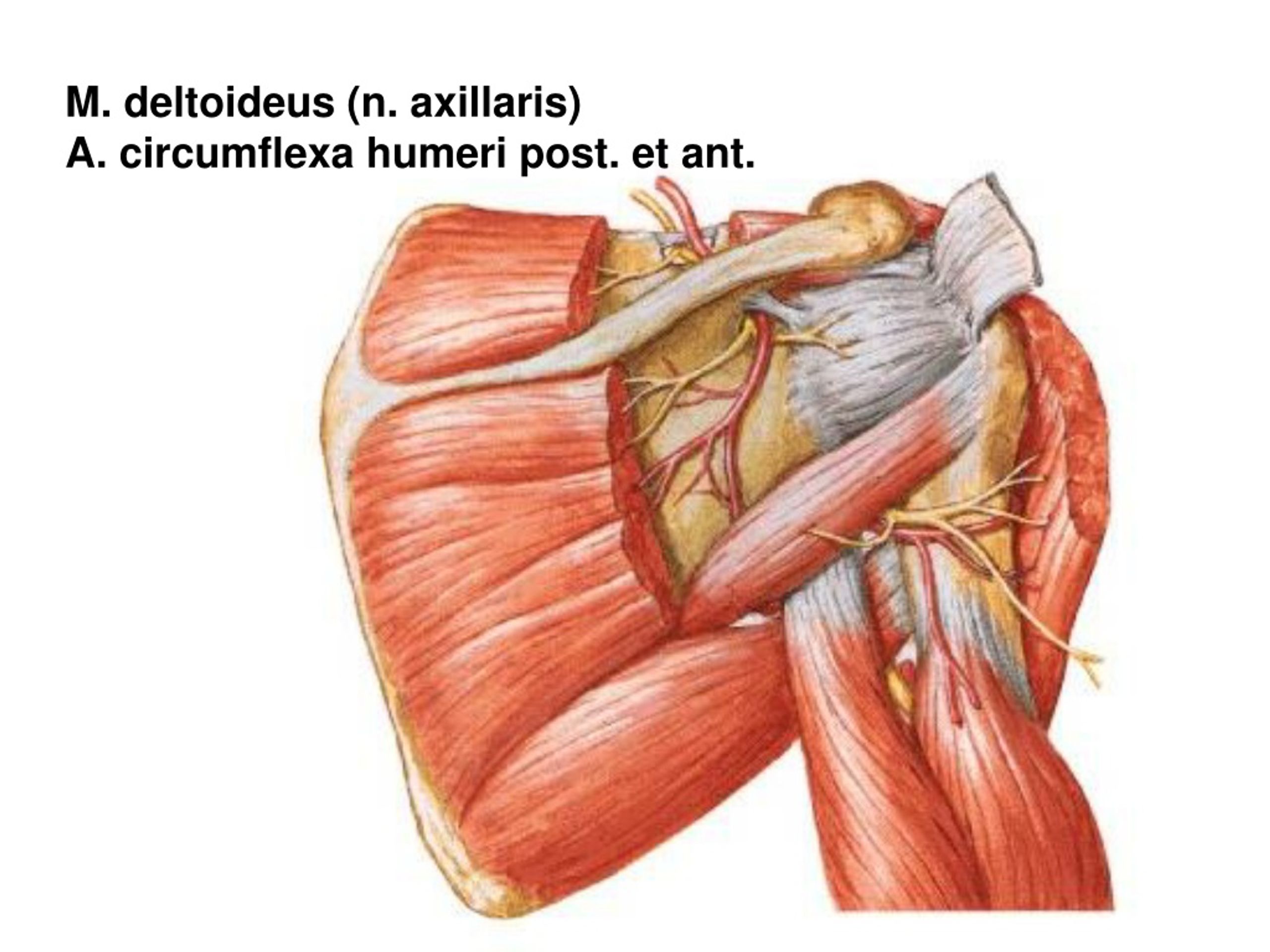
REGIONAL ANATOMY OF THE UPPER LIMB Regio deltoidea
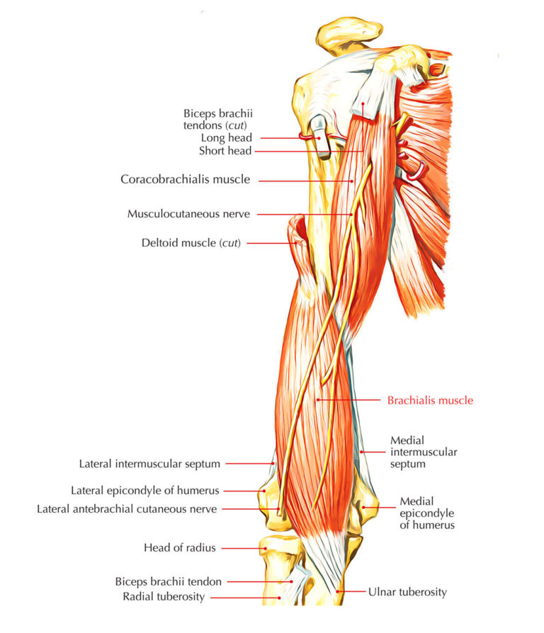
The musculocutaneous nerve emerges as the terminal branch of the lateral cord of the brachial plexus, from the C5-C7 nerve roots.The first muscle it enters is coracobrachialis and gives branches to this muscle before entering it. From here it runs in the flexor compartment superficial to the brachialis but deep to the biceps brachii muscle. As it descends it innervates both of these muscles.
PPT REGIONAL ANATOMY OF THE UPPER LIMB PowerPoint Presentation, free download ID8955187
Regio Brachii Anterior. Course. Anatomy (06) 159 Documents. Students shared 159 documents in this course. University Medical University-Pleven. Info More info. Academic year: 2017/2018. Listed book Gray's Anatomy for Students. Uploaded by: Cristina Ribera. Universitat Rovira i Virgili. 0 followers. 145 Uploads 999+ upvotes. Follow.

Clinical anatomy of the upper limb презентация онлайн
Regio Brachii Batas - batas regio : Proximal: lipat ketiak; Distal : garis penghubung kedua epicondyli humeri; Regio brachii tediri dari regio brachii anterior dan regio brachii posterior. Regio Cubiti Batas: masing - masing 3 jari ke arah proximal dan 3 jari ke arah distal dari garis yang menghubungkan kedua epicondyli. Regio antebrachii.

REGIONAL ANATOMY OF THE UPPER LIMB Regio deltoidea
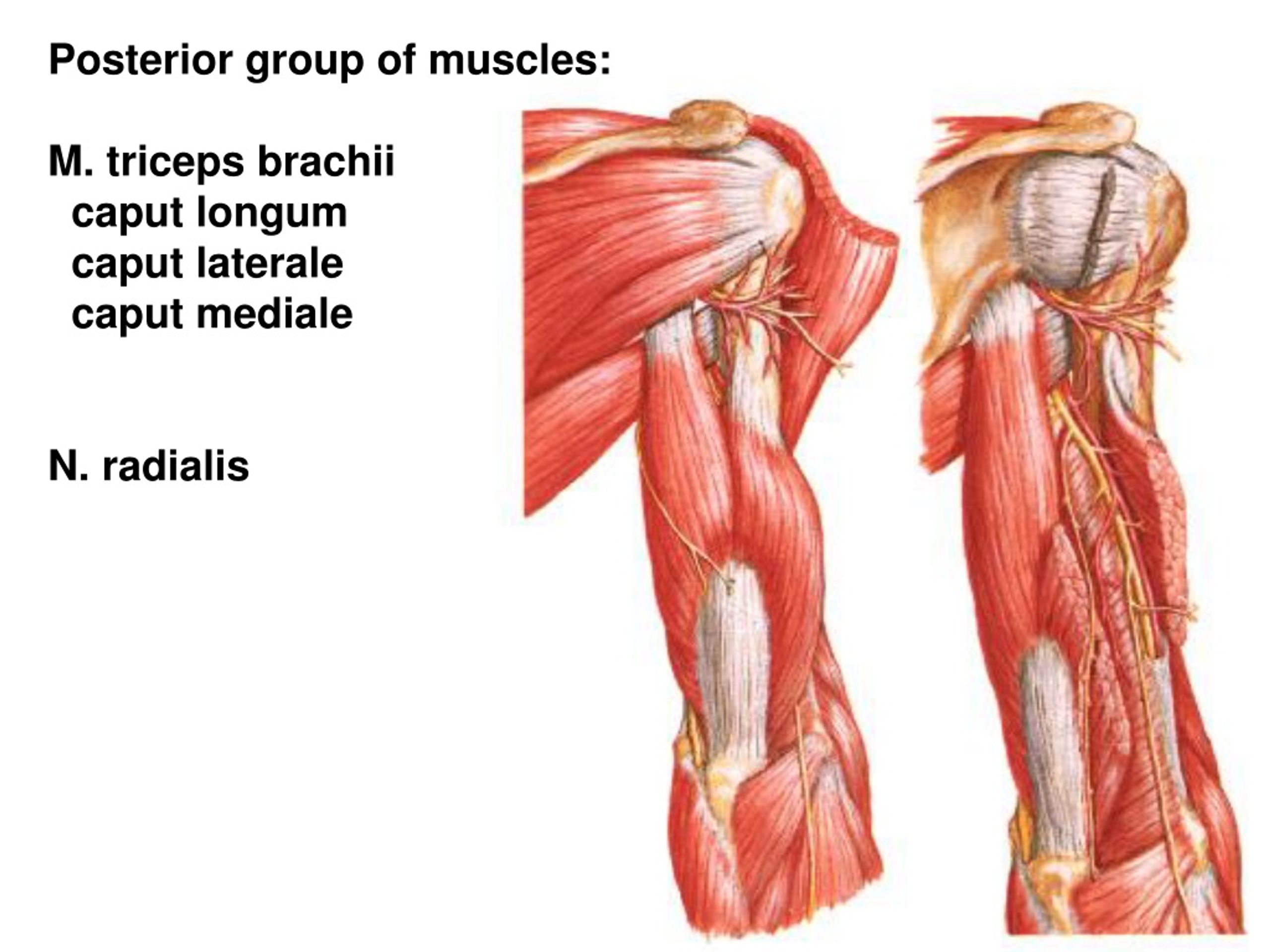
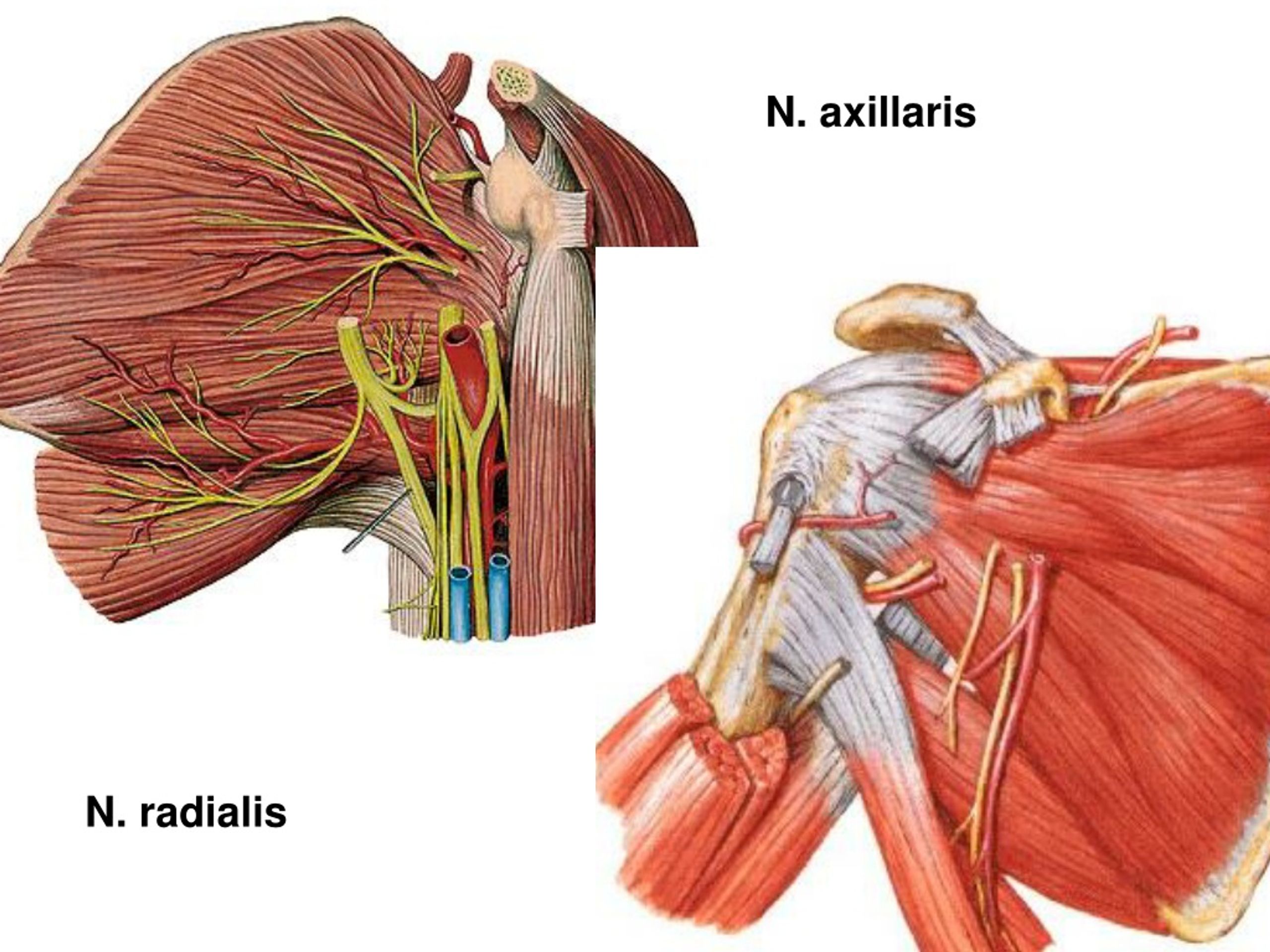
Regio Brachii Posterior. Course: Anatomy (06) 158 Documents. Students shared 158 documents in this course. University: Medical University-Pleven. AI Chat. Info More info. Download. AI Quiz. Save. T OPIC 225. REG IO BRACHII POST ERIOR. 1. LANDMARKS-Deltoid m.-Pe ctor alis major m.-Latissimus dor si m.
Schematic Drawing Showing The Anatomical Landmarks Of The Brachial Images
V. Oberarmstreckseite, Regio brachii posterior. 1. N. radialis Und Begleitgefäße. Der breite sog. Sulcus ni. radialis humeri ist die Ursprungsfläche des lateralen Teiles des M. brachialis, der die Deltoideus- insertion lateral-dorsal umgreift.
PPT REGIONAL ANATOMY OF THE UPPER LIMB PowerPoint Presentation, free download ID8955187
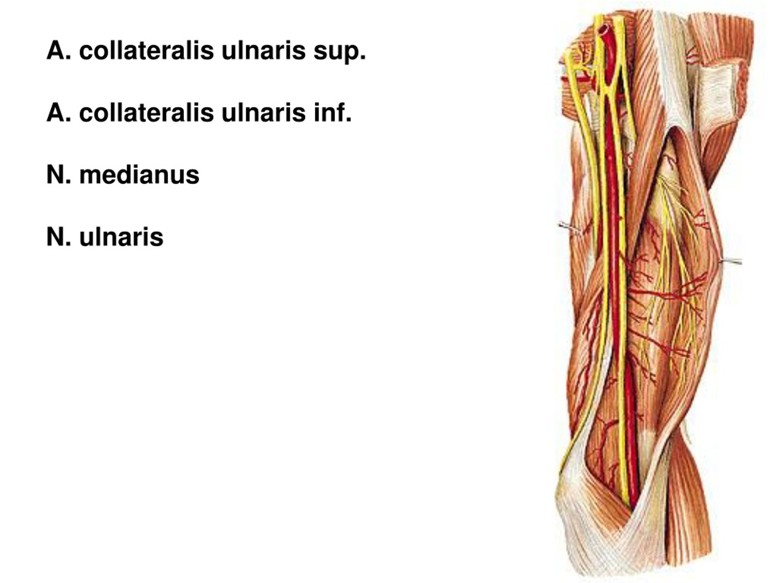
The posterior brachial region, regio brachii posterior. boundaries - behind the humerus: sulcus bicipitalis medialis. sulcus bicipitalis lateralis. surface anatomy: skin - thicker and less mobile. subcutaneous layer - abundant fat and loose connective tissue: n. cutaneus brachii posterior.
Brachialis Muscle Earth's Lab
Regio Brachii 3.4.1. Medial Approach. When the cadaver is positioned in dorsal recumbency, allowing for medial dissection of the upper arm, the pectoral muscles obscure the inspection of the armpit (Figure 5A). Caudally located in the axillary region, ventral to the latissimus dorsi muscle, are the axillary lymph nodes embedded in adipose tissue.
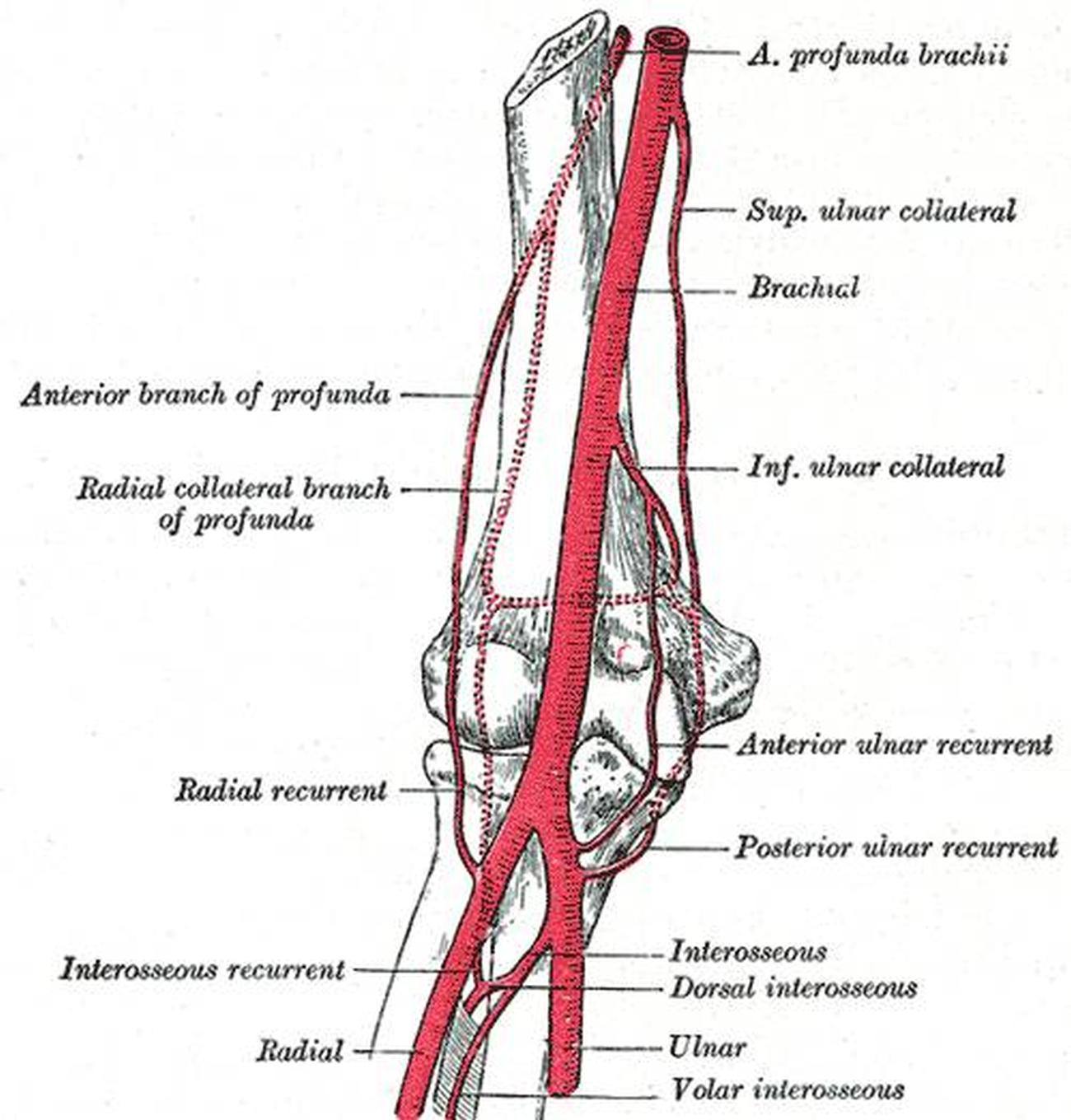
Pictures Of Brachial Artery
Thus, regio brachii anterior et posterior should be changed to regio brachialis anterior et posterior and regio antebrachii anterior et posterior should be changed to regio antebrachialis anterior et posterior. Arteria collateralis media is wrongly referred to as the "medial collateral artery" in the TA.
PPT REGIONAL ANATOMY OF THE UPPER LIMB PowerPoint Presentation, free download ID8955187
a triangle in the upper chest region that is bounded medially by the clavicle, superiorly by the deltoid m. and inferiorly by the pectoralis major m. deltopectoral triangle is pierced by the cephalic vein on its course from the upper limb to join the axillary vein in the axilla. hypothenar compartment. compartment in the hand bounded by the.

PPT REGIONAL ANATOMY OF THE UPPER LIMB PowerPoint Presentation, free download ID8955187
Oberarmbeugeseite, Regio brachii ventralis. 4. Unterbinden der Armschlagader. Heidrich: Bruns' Beitr. 124, 607 (1921). Google Scholar V. Oberarmstreckseite, Regio brachii posterior. 1. N. radialis und Begleitgefäße. Der breite sog. Sulcus ni. radialis humeri ist die Ursprungsfläche des lateralen Teiles des M. brachialis, der die Deltoideus.

profunda_brachii Arm Anatomy, Anatomy Study, Human Anatomy, Hand Therapy, Massage Therapy
The brachial vein (deep vein) accompanies the brachial artery in the region of the arm. It is formed by the unification of the ulnar and radial veins at the elbow. The basilic vein joins the brachial vein and becomes the axillary vein at the inferior border of the teres major muscle. At its terminal part the axillary vein is joined by the.
PPT REGIONAL ANATOMY OF THE UPPER LIMB PowerPoint Presentation, free download ID8955187
Regions of perineum. Regions of upper limb. Axillary region. Deltoid region. Brachial region. Anterior region of arm. Posterior region of arm. Cubital region. Antebrachial region.

Biceps Muscles (Brachii & Brachialis)
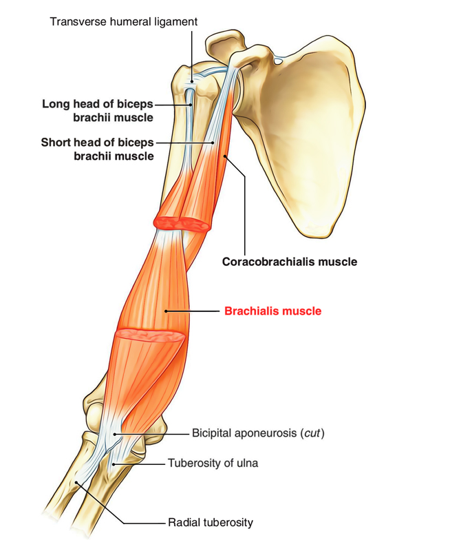
The biceps brachii muscle (biceps) is a large, thick muscle of the arm consisting of two heads.. long head: originates at the supraglenoid tubercle above the glenoid cavity of the scapula.It lies within the intracapsular space but it still remains extrasynovial. The long biceps tendon makes a sharp turn at the humeral head and continues its course in the bicipital groove (intertubercular sulcus).

Oberarm Muscle health, Medical illustration, Biceps brachii
Brachialis muscle (Musculus brachialis) The brachialis muscle is a prime flexor of the forearm at the elbow joint. It is fusiform in shape and located in the anterior (flexor) compartment of the arm, deep to the biceps brachii. The brachialis is a broad muscle, with its broadest part located in the middle rather than at either of its extremities.
Brachial Artery Diagram
Regio brachialis. Latin synonym: Regio brachii Synonym: Arm region Definition. There is no definition for this structure yet. Suggest a definition See the definition in: Français I agree herein to the cession of rights to my contribution in accordance.

Muscles of the Upper Arm Biceps Triceps TeachMeAnatomy
Study with Quizlet and memorize flashcards containing terms like Sebutkan Musculus yang ada di regio brachii ventralis, Apakah yang di maksud dengan lacertus fibrosus, M. Biceps Brachii caput longum bertempat di sisi and more.
