
Discseel Procedure Solusi Degeneratif Tulang Belakang Tagar
Background. Depending on the location of the herniated disc at the shoulder, axilla, or ventral side of the compression nerve root, various puncture sites and channel entrances were selected so that the goal of targeted removal of the herniated disc could be achieved by a full-endoscopic technique.
【Discseel Procedure (DST)】Treatment case of a patient in his 70's, who had undergone
The intervertebral discs are secondary cartilaginous joints, also known as symphyses 7 . Each intervertebral disc is comprised of: peripheral annulus fibrosus. central nucleus pulposus. hyaline cartilage (vertebral side) and fibrocartilage (nucleus pulposus side) Above and below the intervertebral disc are the vertebral body endplates .

Diagnosis Spondylolisthesis Alomedika
Penyempitan Diskus Invertebralis memiliki gejala yang harus anda waspadai. Gejala-gejala tersebut adalah sebagai berikut: Pinggang terasa nyeri. Pinggang bisa saja tiba-tiba terasa nyeri. Ketika anda mengalami hal demikian, anda harus tanggap dalam menentukan penanganan pertama pada pinggang nyeri.
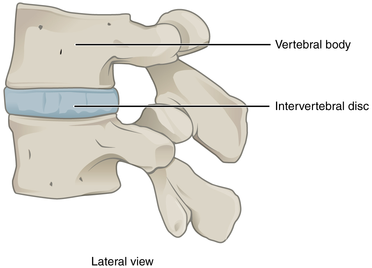
Spine Basics OrthoInfo AAOS
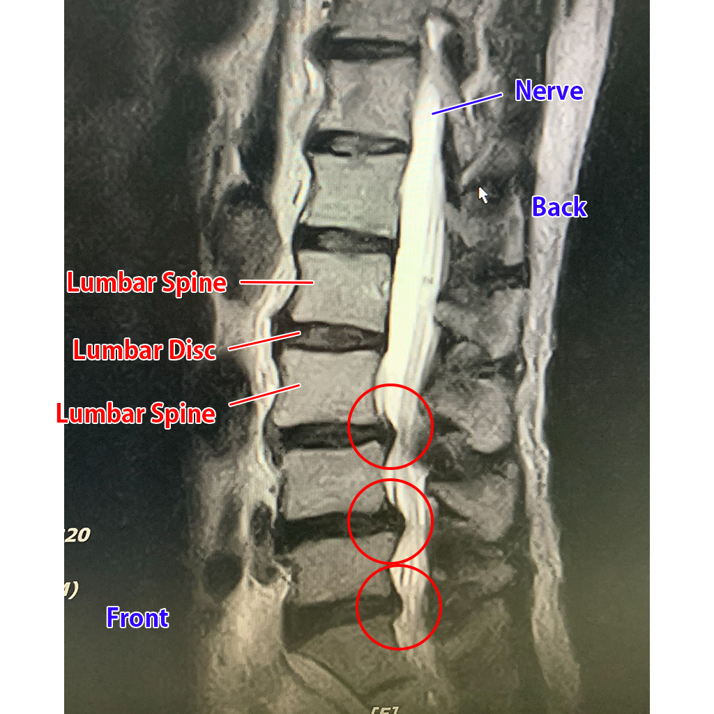
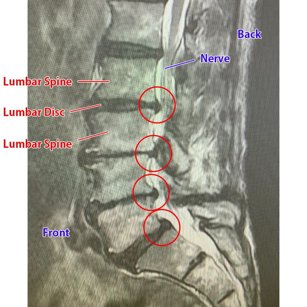
Arti penyempitan "foramen intervertebralis L5-S1" adalah penyempitan antara bantalan tulang -tulang tersebut sehingga jarak antar tulang menjadi lebih rapat, lebih tepat nya pada ruas akhir tulang pinggang. Anda belum tentu mengalami suatu saraf terjepit/HNP. Penyempitan pada celah tulang belakang belum tentu terjadi penjepitan saraf.

Gambar Diskus Intervertebralis PDF
Definition/Description. The intervertebral disc (IVD) is important in the normal functioning of the spine. It is a cushion of fibrocartilage and the principal joint between two vertebrae in the spinal column. There are 23 discs in the human spine: 6 in the cervical region (neck), 12 in the thoracic region (middle back), and 5 in the lumbar.
Intervertebral Discs Anatomy and Embryology Kenhub
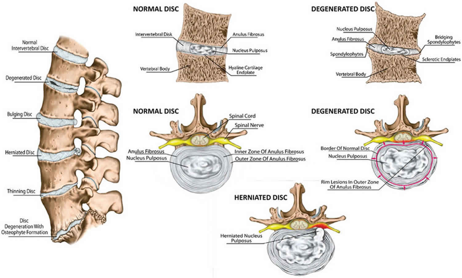
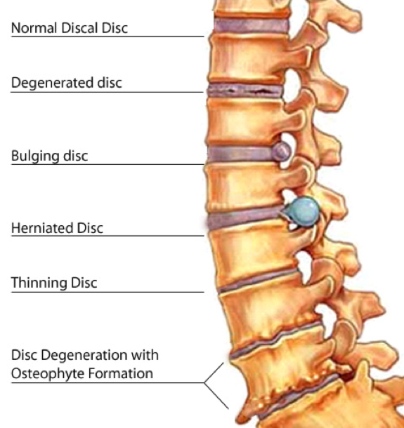
Penyempitan ruang diskus yang terus berlanjut dan terjadinya fibrosis secara bersamaan dengan pembentukan osteofit dan jembatan transdiskal: Tabel 1. Alur degeneratif yang terjadi pada diskus intervertebralis oleh Willis and Bernard (2) Terdapat beberapa faktor risiko untuk kondisi ini, yaitu : (2) Usia; Tingkat aktivitas dan pekerjaan;
Dari A sampai Z tentang Degenerasi Diskus Intervertebra Unair News
CT scan tulang belakang mampu memvisualisasikan tulang belakang secara lebih rinci dan dapat mendiagnosis penyempitan saluran tulang belakang (stenosis tulang belakang) saat ini.. Ahli medis menggunakan MRI untuk memvisualisasikan diskus intervertebralis, termasuk tingkat herniasi diskus jika ada. Selain itu, MRI juga bisa memvisualisasikan.

Laser treatment (PLDD) for Disk Herniation NLC Nonaka Lumbago Clinic
Berikut ini komplikasi spondylosis yang mungkin terjadi adalah: Stenosis tulang belakang. Kondisi penyempitan saluran saraf pada tulang belakang yang menyebabkan gejala mati rasa, kesemutan, atau kelemahan pada kaki. Radikulopati serviks. Perubahan pada cakram atau tulang di punggung yang menyebabkan saraf terjepit, sehingga menimbulkan nyeri.
Intervertebral disc anatomy, function, degeneration, herniation
The intervertebral discs are approximately 7-10 mm thick and 4 cm in diameter (anterior - posterior plane) in the lumbar region of the spine. It consists of a thick outer ring of fibrous cartilage called the anulus (derived from the Latin word "anus" meaning ring) or annulus (anulus fibrosus disci intervertebralis), which surrounds an.
Osteopathic Manual Therapy and Pilates in the Rehabilitation Process for Disc Herniation and
Calendars and Case Information View case summaries, defendant and plaintiff names, and case information. Civil, Criminal, Family, and Probate Case Search Calendar Search Calendars for individual judges and programs in the Civil, Criminal, and Probate Divisions can be accessed through the following link: Civil, Criminal, and Probate What you need to know about Sealed Court Records By

Diskus Intervertebralis Sintetik Mulai Dicoba pada Hewan
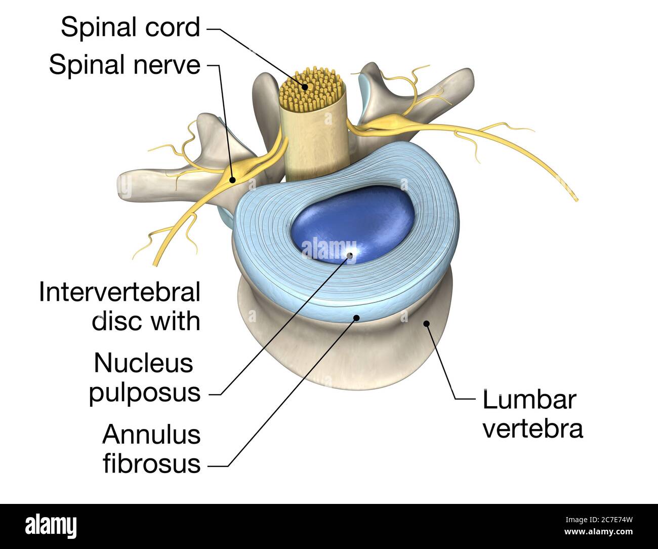
An intervertebral disc is a structure located between adjacent vertebrae of the spine. It consists of a tough outer layer called the annulus fibrosus and a gel-like center called the nucleus pulposus. The intervertebral disc acts as a cushion, allowing the spine to bend and twist without damaging the vertebrae.
3D illustration showing lumbal vertebra with intervertebral disc, medically 3D illustration
Diskus intervertebralis terdiri dari dua struktur utama, yaitu nukleus pulposus (NP) dan annulus fibrosus (AF).. penyempitan diskus, degenerasi musinosa, gas intradiskus (vakum), osteofit, perubahan inflamasi, dan sklerosis subkondral. Fisura anulus merupakan predisposisi kelemahan, yang memungkinkan material diskus menonjol atau bermigrasi.
Classification of Joints · Anatomy and Physiology
Tanda spondylosis serviks, penyempitan foramina intervertebralis, osteofit, dan perubahan degeneratif lainnya merupakan hal yang lazim pada orang dengan dan tanpa nyeri leher.. Degenerasi diskus dapat menyebabkan hilangnya ketinggian antara vertebra, menempatkan kekuatan kompresi yang lebih besar pada sendi facet posterior..
【Discseel Procedure (DST)】Treatment case of a patient in his 60s that suffered for more than 20
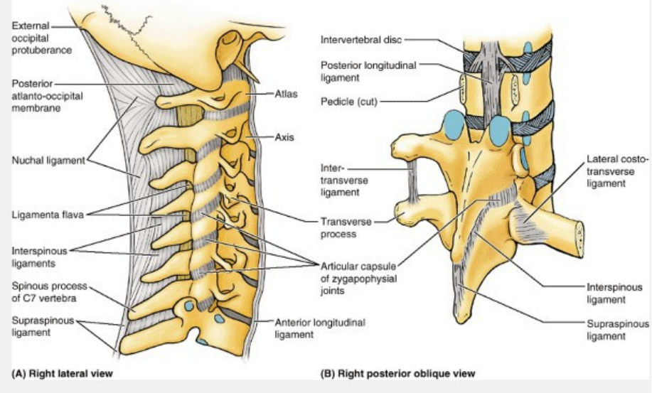
Synonyms: none. The intervertebral joints connect directly adjacent vertebrae of the vertebral column. Each intervertebral joint is a complex of three separate joints; an intervertebral disc joint (intervertebral symphysis) and two zygapophyseal (facet) joints. This article will describe the anatomy and function of the intervertebral joints.

Diskus intervertebralis II Anatomi og Fysiologi
This review article describes anatomy, physiology, pathophysiology and treatment of intervertebral disc. The intervertebral discs lie between the vertebral bodies, linking them together. The components of the disc are nucleus pulposus, annulus fibrosus and cartilagenous end-plates. The blood supply to the disc is only to the cartilagenous end.
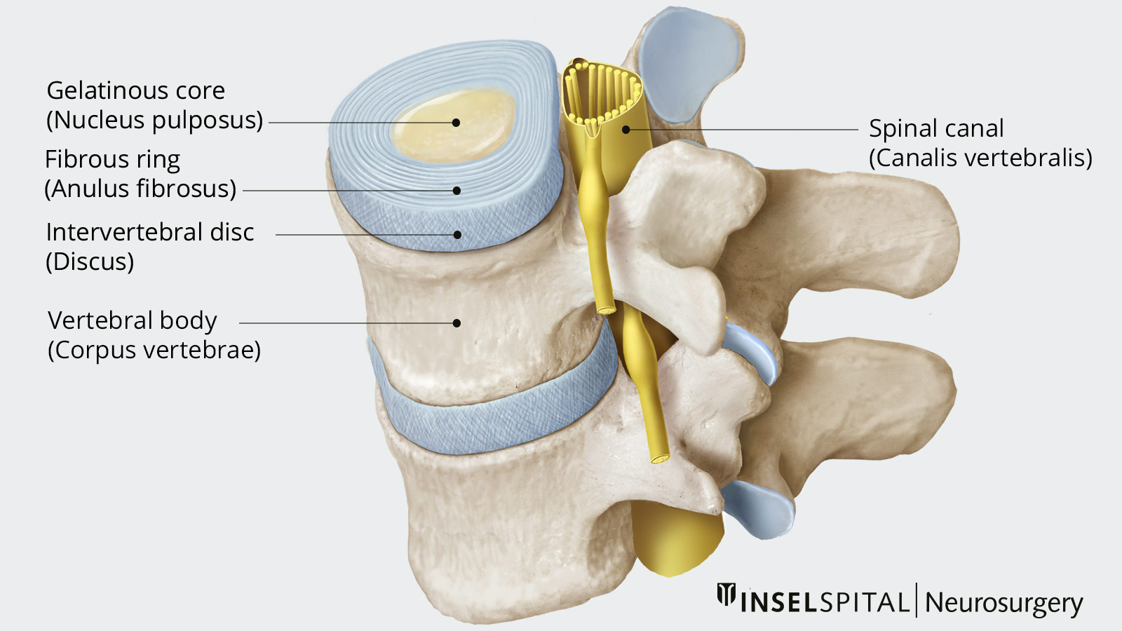
Herniated Disc Neurosurgery Inselspital Bern
Adjacent vertebrae articulate through zygapophyseal joints between the respective superior and inferior facets of the vertebral articular processes as well as through the joints of the vertebral bodies. While the former serves to limit the spine's range of motion, the latter increases it and provides the majority of the spine's weight-bearing capacity. The inferior surface of the superior.
