
Avulsion fracture of the calcaneus CMAJ
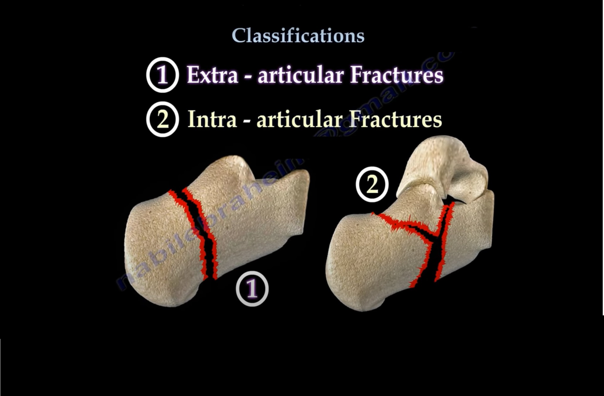
The calcaneus is the most commonly fractured tarsal bone and accounts for about 2% of all fractures 2 and ~60% of all tarsal fractures 3. Pathology. Calcaneal fractures can be divided broadly into two types depending on whether there is articular involvement of the subtalar joint 2,7,8: extra-articular: 25-30%. anterior calcaneal process fracture
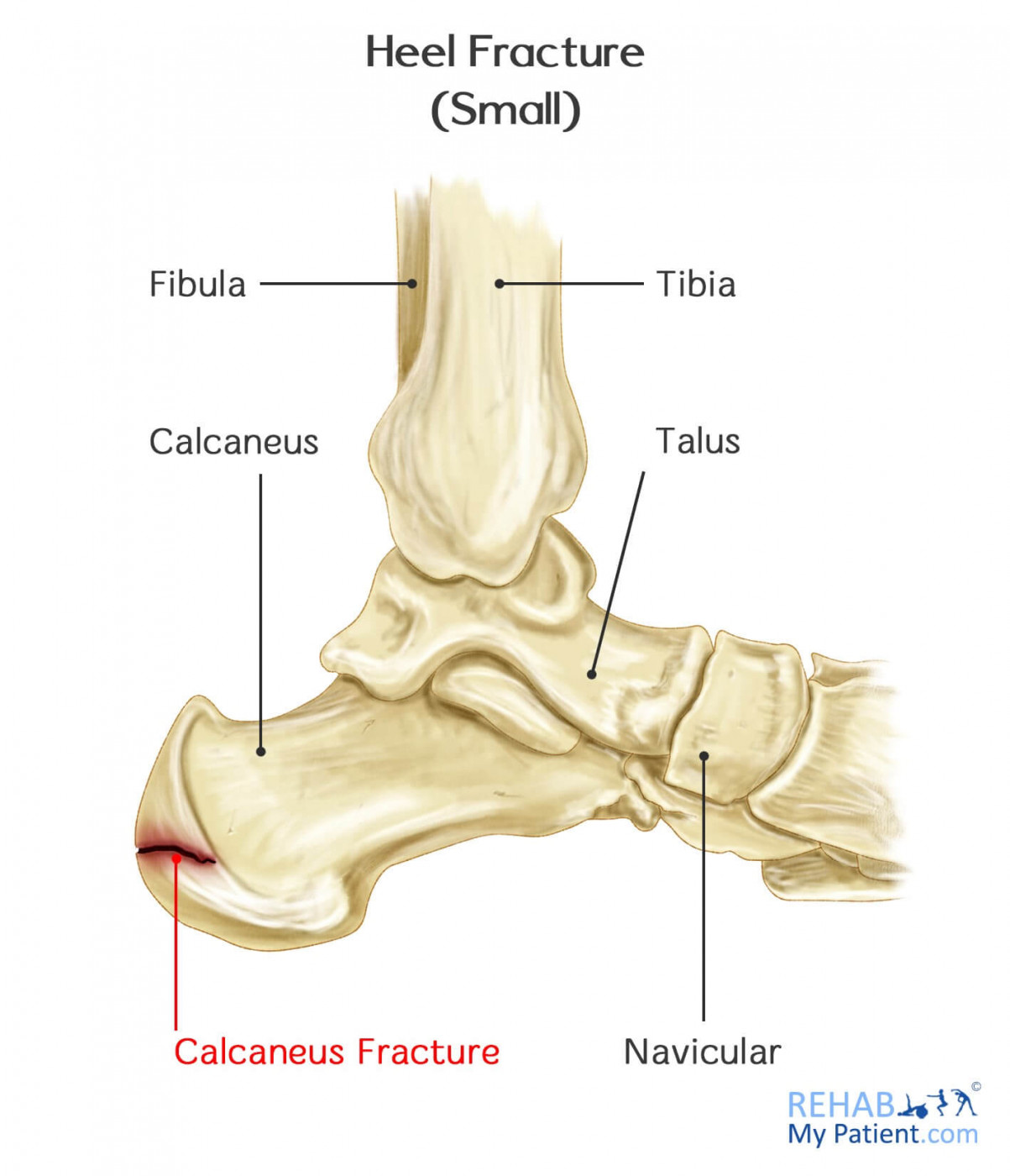
Heel Fracture Rehab My Patient
Fraktur kalkaneus adalah cedera serius tetapi jarang terjadi. Mereka hanya menyumbang 1 dan 2% dari semua patah tulang. Namun, jika tidak didiagnosis dan diobati segera, mereka dapat menyebabkan kecacatan jangka panjang. Hingga 10% dari fraktur ini tidak dikenali pada kunjungan awal ke kamar darurat.

Fracture of the Calcaneus Trauma Case Studies CTisus CT Scanning
Fraktur calcaneus paling sering adalah pada badan calcaneus. Fraktur pada prosesus anterior kira-kira 10 - 15% daripada cedera ekstra-artikular, dan merupakan jenis fraktur calcaneus yang paling sering terjadi pada wanita dibanding laki-laki.6 Fraktur intra-artikular mewakili hampir 70% kejadian pada orang dewasa.
Fractured calcaneal spur. radRounds Radiology Network
Laporan Pendahuluan Fraktur pengertian fraktur adalah pemisahan atau patahnya tulang (doenges, fraktur adalah patahnya tulang yang biasanya disebabkan oleh. Skip to document. Ask AI.. FRAKTUR: CALCANEUS. B. KLASIFIKASI. Fraktur dapat diklasifikasikan dalam dua jenis klasifikasi, yaitu menurut kondisi permukaan kulit dan yang kedua menurut.

FileCalcaneus Fracture.jpg Wikipedia
Calcaneal fractures can be divided into two groups: intra-articular and extra-articular calcaneal fractures. Intra-articular fractures have a lower prognosis. To determine the kind of fracture and if there is a fracture, medical imaging is needed. The rehabilitation program consists of 3 stages postoperatively and is very important to enhance.
Avulsion Fractures Of Talus And Calcaneus Image Radiopaedia Org My XXX Hot Girl
Fraktur dengan kulit tetap utuh disekitar fraktur tidak menonjol keluar dari kulit dan tidak terdapat hubungan antara fragmen tulang dengan dunia luar adalah fraktur tertutup. Jaringan akan mengalami kerusakan pada kanalis havers dan jaringan lunak diarea terjadinya fraktur, selanjutnya akan terbentuk bekuan darah dan benang-benang fibrin serta.
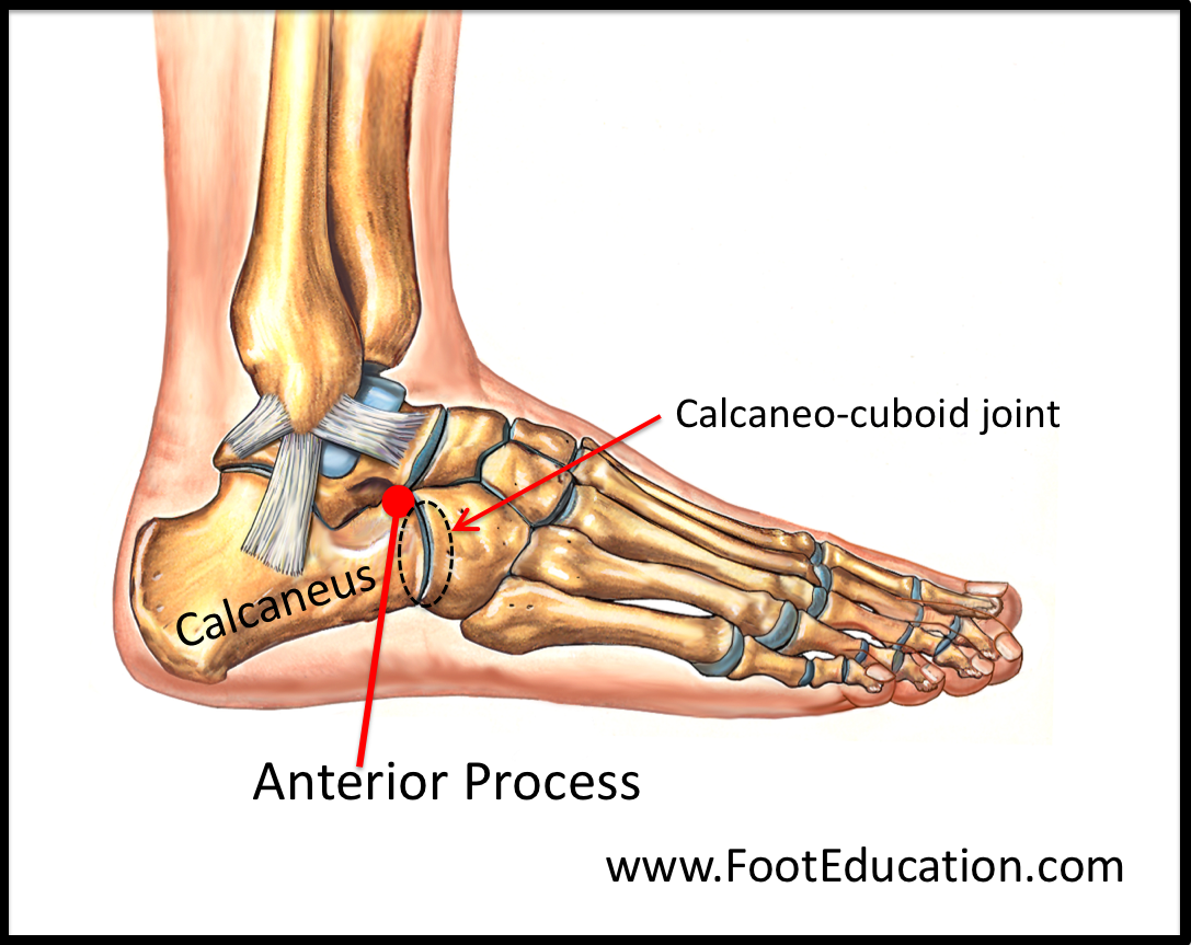
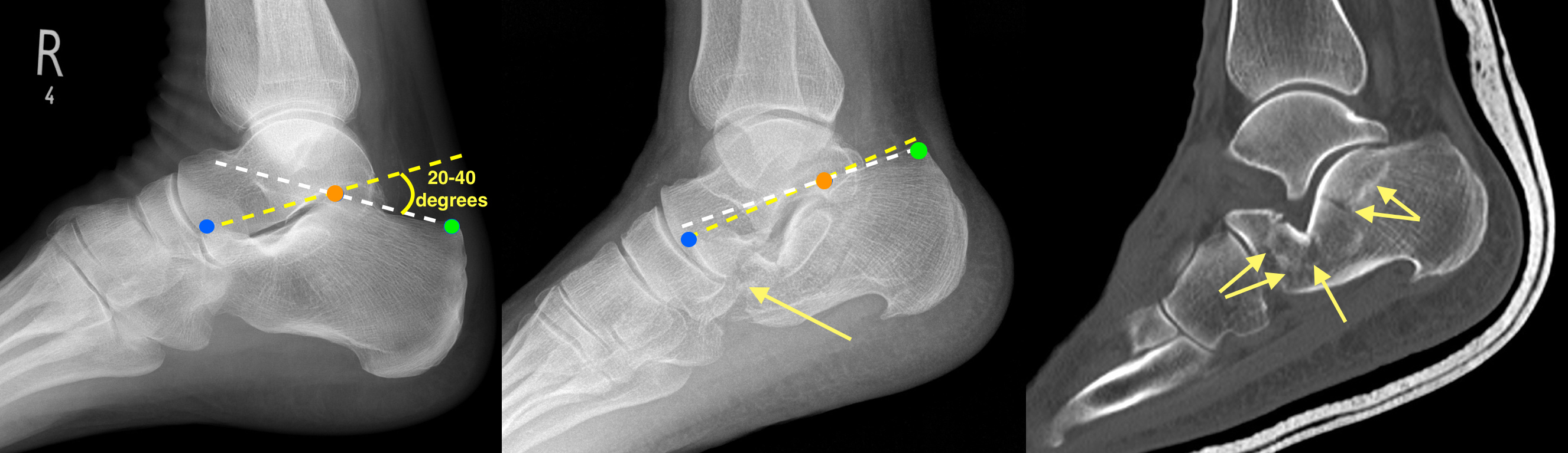
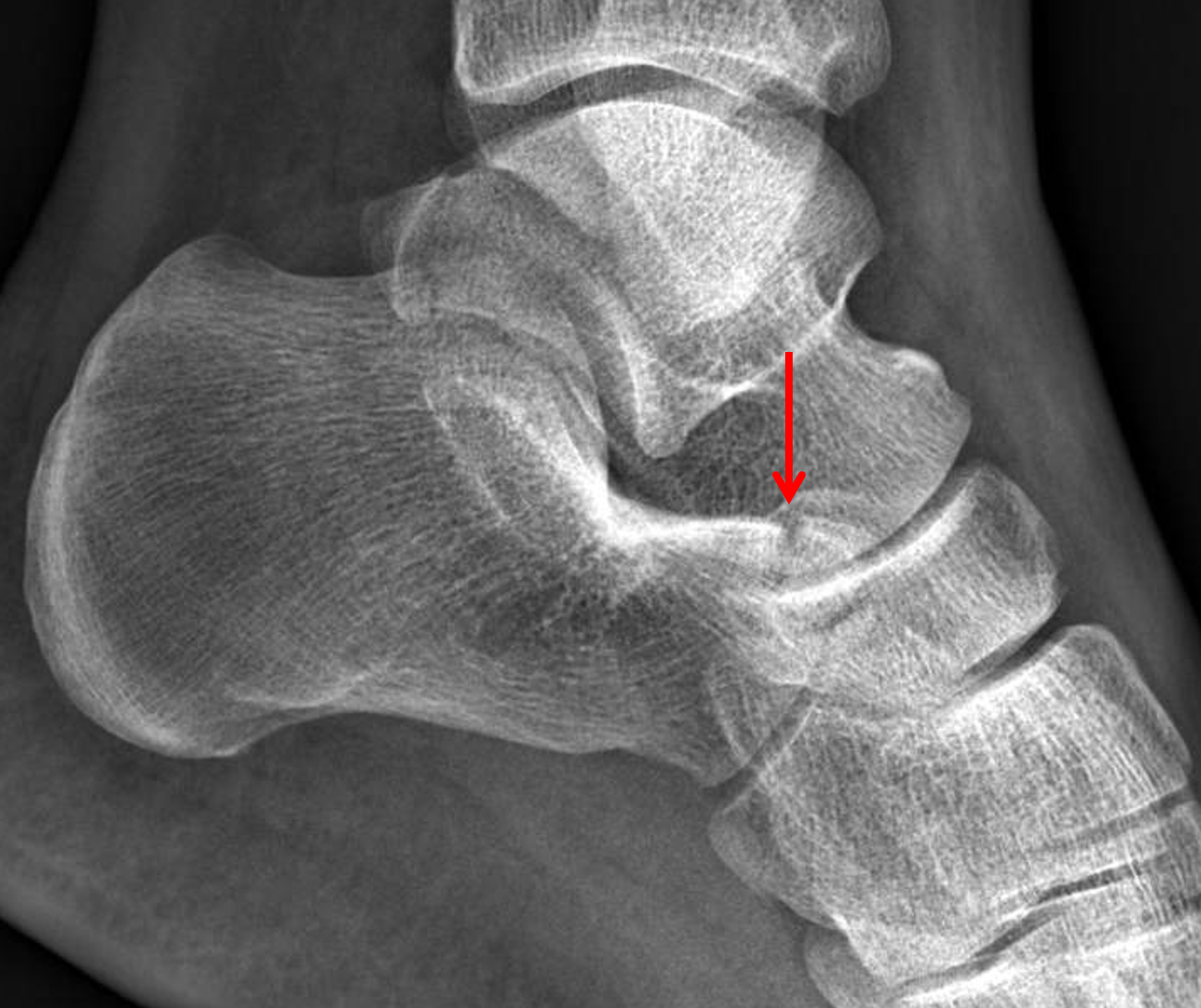
Anterior Process Fracture of the Calcaneus FootEducation
Tujuannya adalah agar pasien dapat kembali ke fungsi atletik seperti sebelumnya. Pencegahan Calcaneal Bursitis. Nyeri dapat dihindari bila pasien segera menghentikan aktivitasnya bila mulai melihat dan merasakan tanda-tanda adanya bursitis. Calcaneal bursitis dapat dicegah dengan melakukan hal-hal berikut ini: Lakukan gerakan olahraga dengan benar.
AVULSION FRACTURE OF THE CALCANEAL TUBEROSITY
The treatment of displaced fractures of the talus and calcaneus is associated with a considerable learning curve. Malunion results in significant limitations of global foot function and painful posttraumatic arthritis. While early reduction of dislocations and fracture dislocations represent an emergency situation, the timing of definitive fixation has no measurable impact on the results and.
Avulsion fracture of the calcaneus CMAJ
Calcaneal Fracture. Muhammad Fadly. I PENDAHULUAN Fraktur calcaneus adalah fraktur paling sering pada os tarsal. Penatalaksanaannya fraktur ini sukar disebabkan kerana jenis fraktur yang bervariasi dan seringnya terjadi komplikasi. Fraktur calcaneus dianggap kasus yang menantang pada ahli ortopedik disebabkan karena rendahnya kepuasan pasien.
Calcaneal Fractures Part 1 —
Calcaneus fractures are rare but potentially debilitating injuries. The calcaneus is one of seven tarsal bones and is part of the hind-foot which includes the calcaneus and the talus. The hindfoot articulates with the tibia and fibula creating the ankle joint. The subtalar or calcaneotalar joint accounts for at least some foot and ankle dorsal/plantar flexion. Calcaneal anatomy is demonstrated.
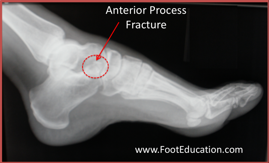
Anterior Process Fracture of the Calcaneus FootEducation
The calcaneus is the most commonly fractured tarsal bone, representing 60 percent of all tarsal fractures in adults [ 1 ]. The peak incidence occurs in younger males [ 2 ]. Most calcaneal fractures are occupational, and are caused by axial loading from a fall [ 2 ]. The majority are displaced intraarticular fractures (60 to 75 percent) [ 2 ].
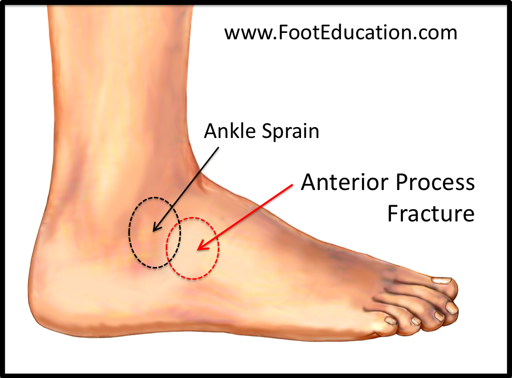
Anterior Process Fracture of the Calcaneus FootEducation
Fraktur calcaneus adalah fraktur paling sering pada os tarsal. Fraktur calcaneus biasanya disebabkan oleh cedera pergelangan kaki yang berputar atau lebih sering akibat terjatuh dari ketinggian, kecelakaan mobil, pergelangan kaki keseleo, penggunaan berlebihan atau stress berulang pada tulang tumit. Fraktur ini
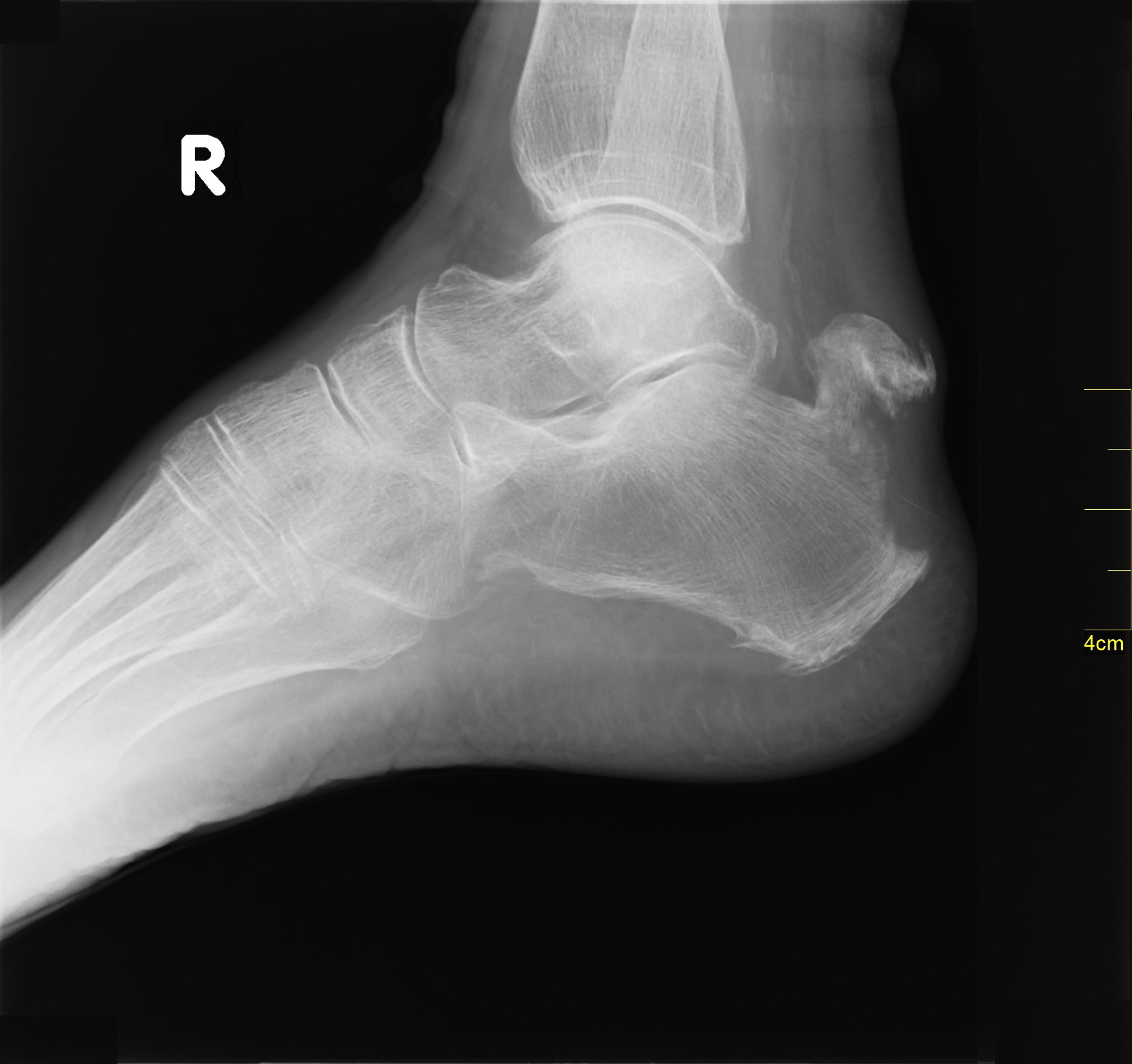
Calcaneal Fracture Radiology at St. Vincent's University Hospital
Calcaneus (Heel Bone) Fractures. A fracture of the calcaneus, or heel bone, can be a painful and disabling injury. This type of fracture commonly occurs during a high-energy event — such as a car crash or a fall from a ladder — when the heel is crushed under the weight of the body. When this occurs, the heel can widen, shorten, and become.

Calcaneus Fractures Dr Ben Beamond Adelaide
Pengertian fraktur (fraktura) atau patah tulang adalah kondisi ketika tulang menjadi patah, retak, atau pecah sehingga mengubah bentuk tulang. Tulang yang mengalami fraktur dapat terjadi di area tubuh manapun. Namun, kasus ini lebih sering terjadi di beberapa bagian tubuh. Misalnya, patah tulang selangka atau bahu, patah tulang tangan (termasuk.
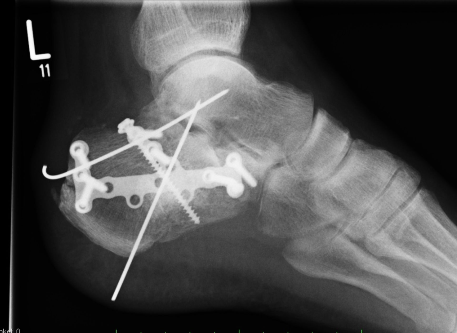
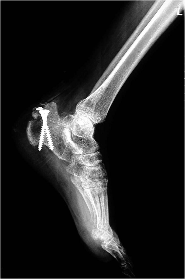
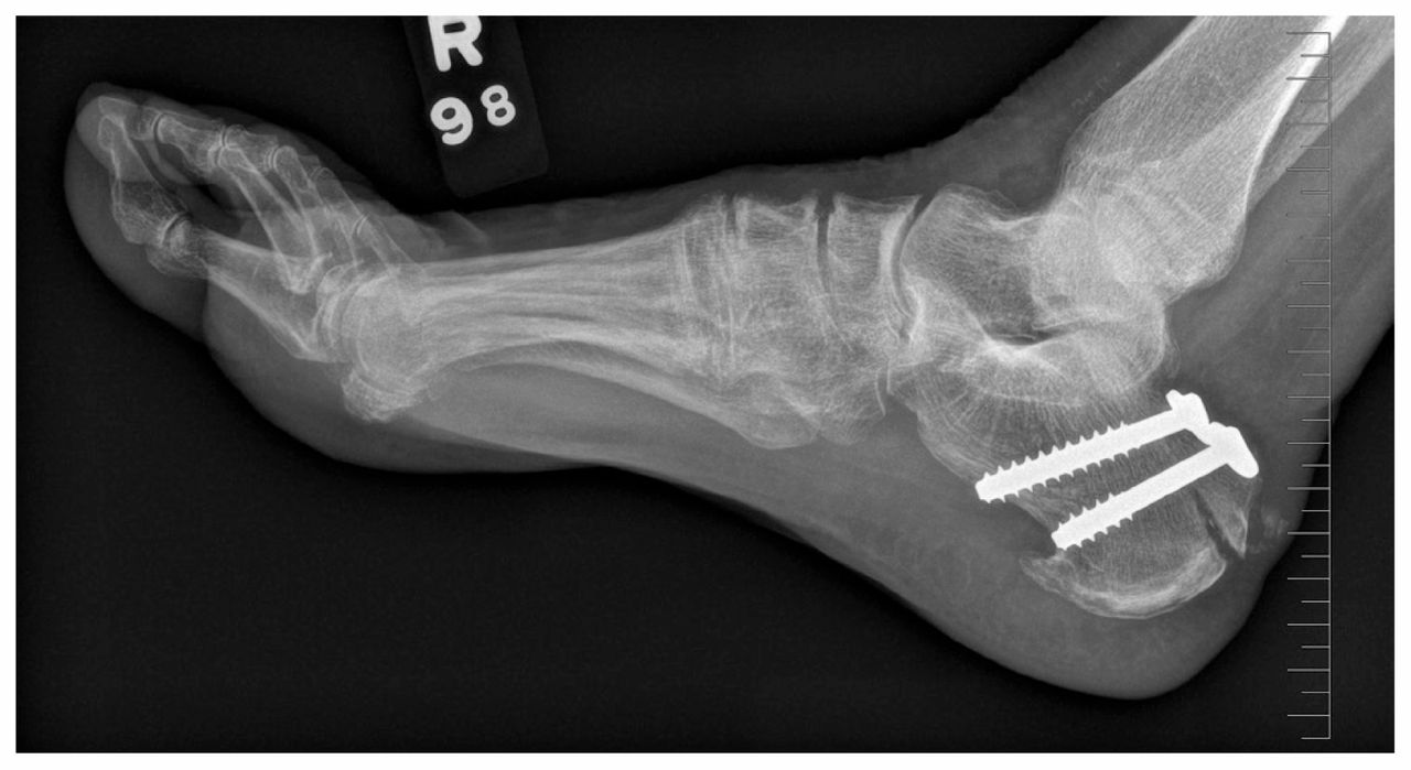
Calcaneusfraktur linksOsteosynthese DocCheck
Briefly, the studies confirmed that the outer layer of soft tissue is the most prone to infection, probably because blood is supplied to the lateral calcaneus solely by the lateral calcaneal artery and, thus, is more likely to develop hypoxia after injury. 22 In addition, the lateral calcaneus has a soft-tissue structure that is thinner than.
Anterior Calcaneus Fracture
Calcaneus Fractures. Calcaneus fractures are the most common fractured tarsal bone and are associated with a high degree of morbidity and disability. Diagnosis is made radiographically with foot radiographs with CT scan often being required for surgical planning.
