
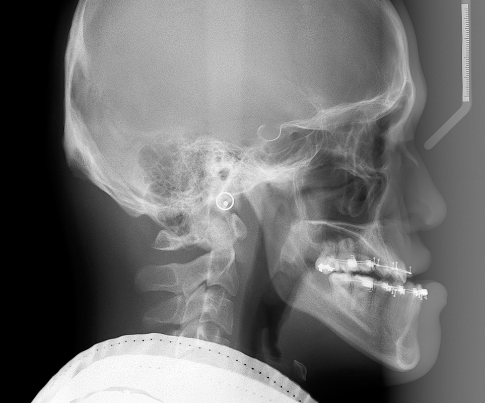
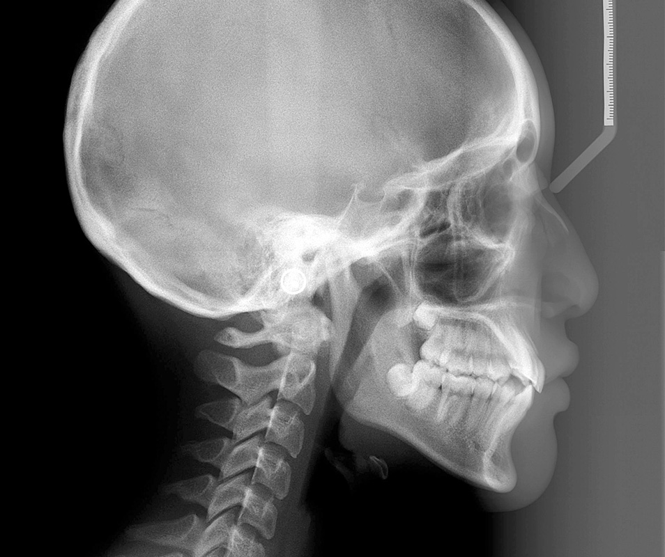
Lateral Cephalometric Radiograph
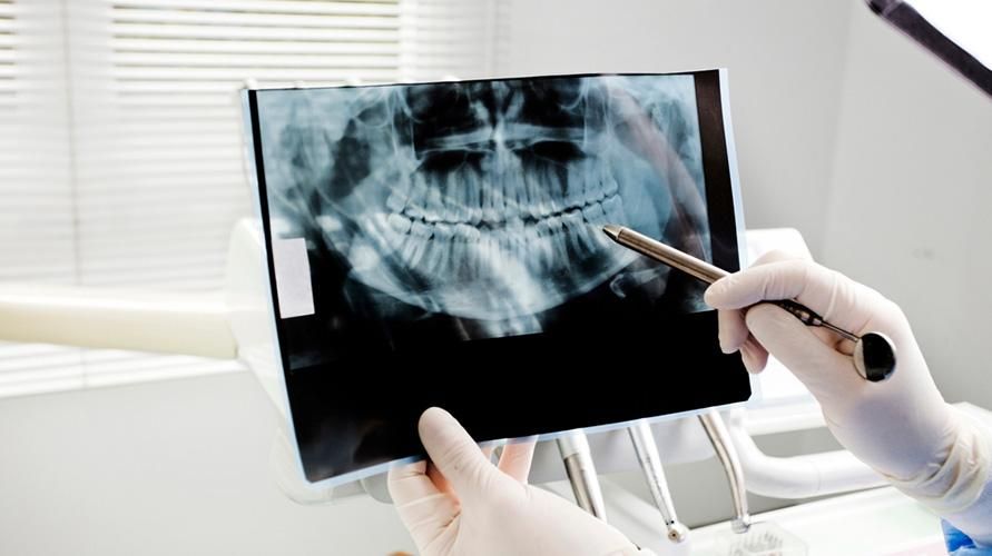
Anda juga akan menjalani prosedur ini ketika merencanakan perawatan gigi palsu, kawat gigi, cabut gigi, dan implan gigi. 2. Cephalometric X-ray. Rontgen gigi ini diambil dari seluruh sisi kepala. Umumnya, dokter melakukan tes pencitraan ini untuk melihat struktur gigi yang berkaitan erat dengan tulang rahang atau fitur wajah.
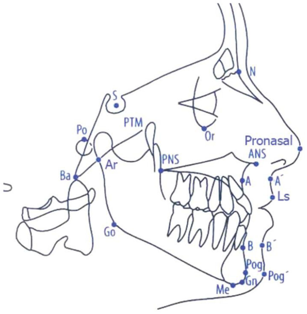
Cephalometric Landmarks Focus Dentistry
Biaya untuk melakukan Rontgen gigi bervariasi, tergantung dari teknik yang dilakukan dan rumah sakit yang menyelenggarakannya. Di rumah sakit swasta di Indonesia, biaya prosedur ini dapat dimulai dari Rp. 65.000 hingga lebih dari Rp. 200.000.
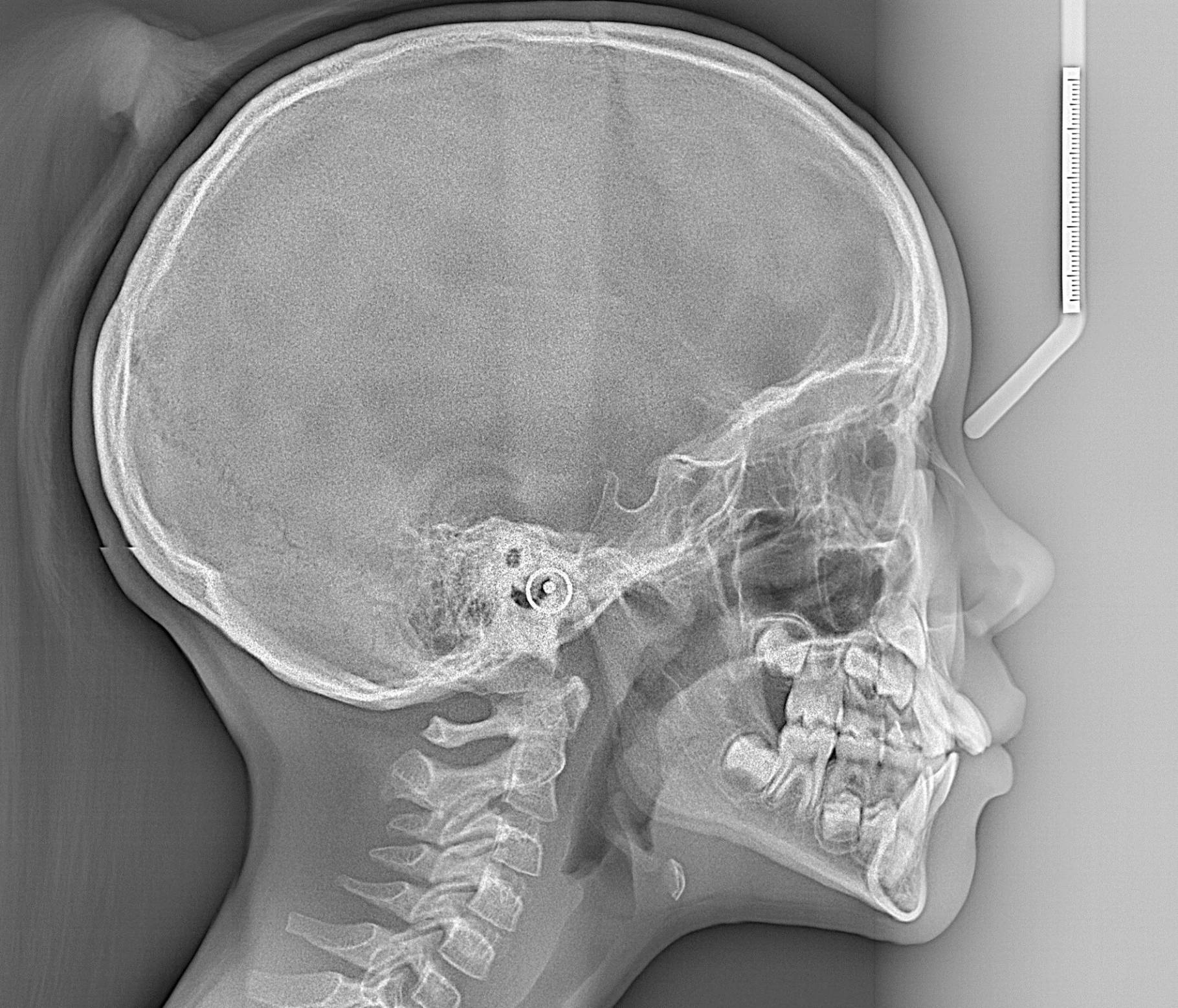
Lateral Cephalometric Radiograph
A cephalometric X-ray, which is also sometimes referred to simply as a ceph, is a diagnostic radiograph used primarily for orthodontic treatment planning. A cephalometric X-ray is taken during the orthodontic records appointment. Cephalometric X-rays are also used by otolaryngologists—doctors who specialize in the treatment of ear, nose, and.
Biaya Rontgen Gigi Panoramic Cephalometric Terbaru Terbaru Biaya.Info
Third, cephalometric tracing with 42 landmark points detection, performed on real and synthesized images by two expert orthodontists, showed consistency with mean difference of 2.08 ± 1.02 mm.

(PDF) Lateral cephalometric analysis for treatment planning in orthodontics based on MRI
Cephalometric x-rays (also called ceph x-rays or radiographs) show a side view of your head, exposing teeth, jaw, and surrounding structures. This technology is considered safe and often useful or necessary to help professionals evaluate and assist patients. This specific type of x-ray is used in diagnosis and treatment planning.
Cephalometric Projection Diagnostic Radiographs Orbit Imaging
Third, cephalometric tracing with 42 landmark points detection, performed on real and synthesized images by two expert orthodontists, showed consistency with mean difference of 2.08 ± 1.02 mm.

Lateral cephalometric radiograph showing the identified landmarks and... Download Scientific
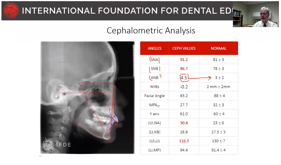
Cephalometric analysis. Cephalometric analysis can be used to analyze the facial skeleton, generally in a two-dimensional (2D) fashion and based on specialized lateral and anteroposterior (AP) skull radiographs (cephalograms). The technique was originally introduced by Broadbent, and since that time, many more complex analytical techniques have.

Cephalometric Analysisi Ricketts Points and Planes Dental anatomy, Dental hygiene student
Cephalometric analysis is an essential tool used in orthodontic diagnosis and treatment planning. The main objectives of correct cephalometric analysis include resolving anteroposterior and vertical maxillary and mandibular base discrepancies. For a diagnostic tool to be of value, it should be precise, reliable and reproducible. Unfortunately, according to some studies, the accuracy of input.
cephalometric analysis Discover Ortho
On comparing the linear cephalometric and photographic variables for the skeletal class II subjects we found the cephalometric parameters convexity (in mm), Witts, mandibular body length had significant p-value indicating that the difference between these photographic and cephalometric parameters was significant and hence the photographic.

Cephalometric Xray of Orthodontic Sherway Gardens Dental Centre
The aim of this study was to validate geometric accuracy and in vivo reproducibility of landmark-based cephalometric measurements using high-resolution 3D Magnetic Resonance Imaging (MRI) at 3 Tesla.
Cephalometric Projection Diagnostic Radiographs Orbit Imaging
4 Retraksi gigi anterior dan perbaikan inklinasi insisif merupakan upaya yang dilakukan untuk mencapai overjet dan overbite yang normal, mengurangi kecembungan wajah, membuat bibir dapat menutup.

Assessment of automatic cephalometric landmark identification using artificial intelligence
A cephalometric analysis can provide a deeper view into the support system of facial esthetics that can assist in planning restorative treatment and predicting the result. Figure 1 Figure 2. Hidden underneath the facial profile photo lies another world of information that may help you with important details for your treatment planning.

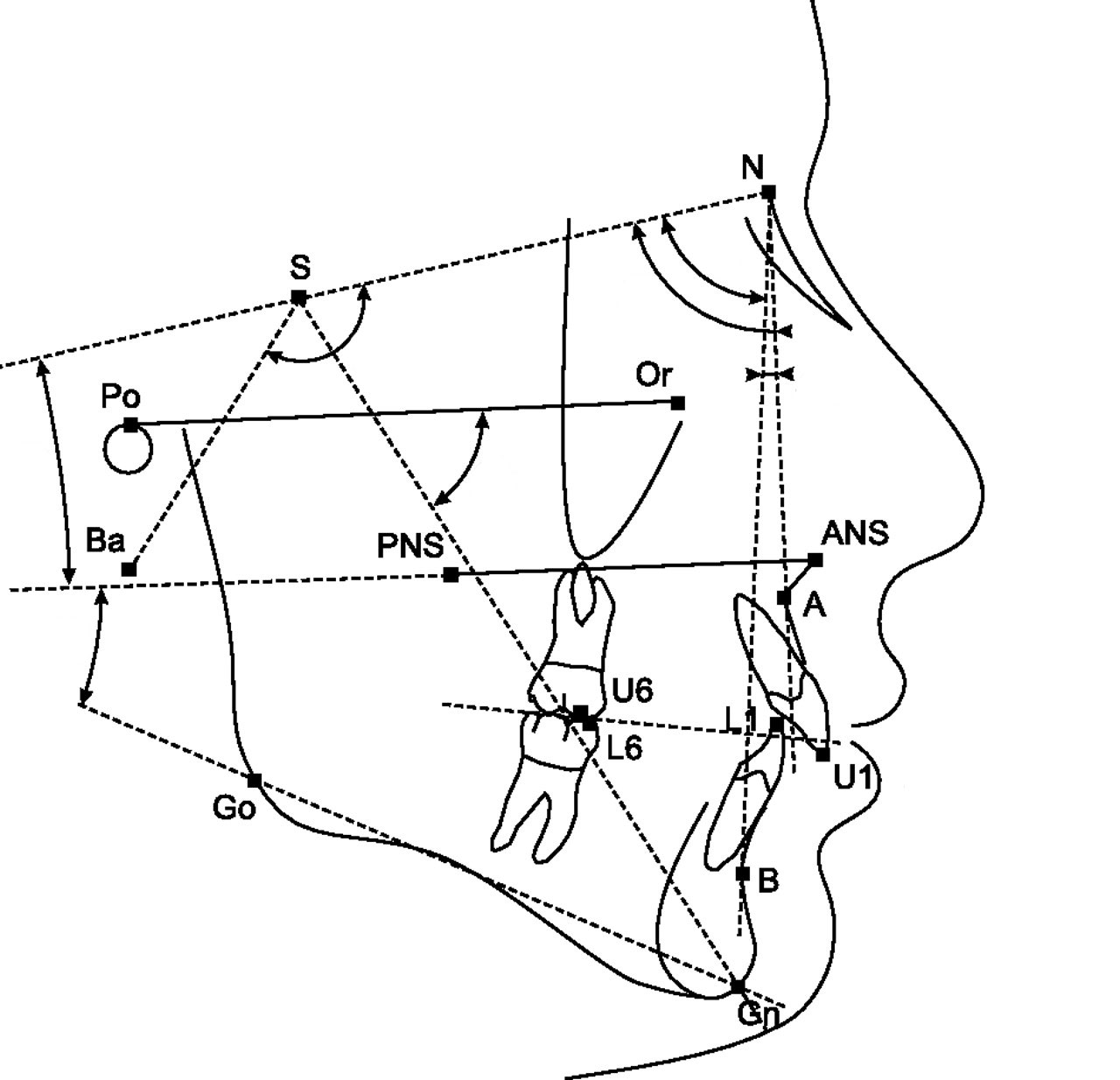
a. Cephalometric soft tissue and skeletal measurements used in the... Download Scientific Diagram
Cephalometric analysis is the clinical application of cephalometry.It is analysis of the dental and skeletal relationships of a human skull. It is frequently used by dentists, orthodontists, and oral and maxillofacial surgeons as a treatment planning tool. Two of the more popular methods of analysis used in orthodontology are the Steiner analysis (named after Cecil C. Steiner) and the Downs.

Angular and linear cephalometric measurements and accepted normal values. Download Scientific
Cephalometric analysis evaluates lateral skull radiographs obtained with a cephalostat to help determine the skeletal pattern and assess treatment difficulty. Cephalometric analysis is indicated when anteroposterior movement is planned but is not required for all orthodontic treatments. The use of cephalometric analysis is justified when the incisor position will be significantly modified.

Initial lateral cephalometric radiograph, tracing, and panoramic... Download Scientific Diagram
Cephalometric radiography is a standardized and reproducible form of skull radiography used extensively in orthodontics to assess the relationships of the teeth to the jaws and the jaws to the rest of the facial skeleton. Standardization was essential for the development of cephalometry - the measurement and comparison of specific points.
Cephalometric Analysis Based on Xray Scans CephX
Corpus ID: 76525813; Determination of final occlusal vertical dimension by cephalometric analysis @inproceedings{Morais2015DeterminationOF, title={Determination of final occlusal vertical dimension by cephalometric analysis}, author={Eduardo Christiano Caregnatto de Morais and B{\'a}rbara Pick Ornaghi and Ana Paula Sponchiado and Jo{\~a}o C{\'e}sar Zielak and Rog{\'e}rio Goulart da Costa and M.