ppt kritisi dan evaluasi radiograf Appendicografi
Pediatric Appendicitis. 2017 Feb;97 (1):93-112. doi: 10.1016/j.suc.2016.08.009. Appendicitis is one of the most common surgical pathologies in children. It can present with right lower quadrant pain. Scoring systems in combination with selective imaging and surgical examination will diagnose most children with appendicitis.
Appendicitis Article
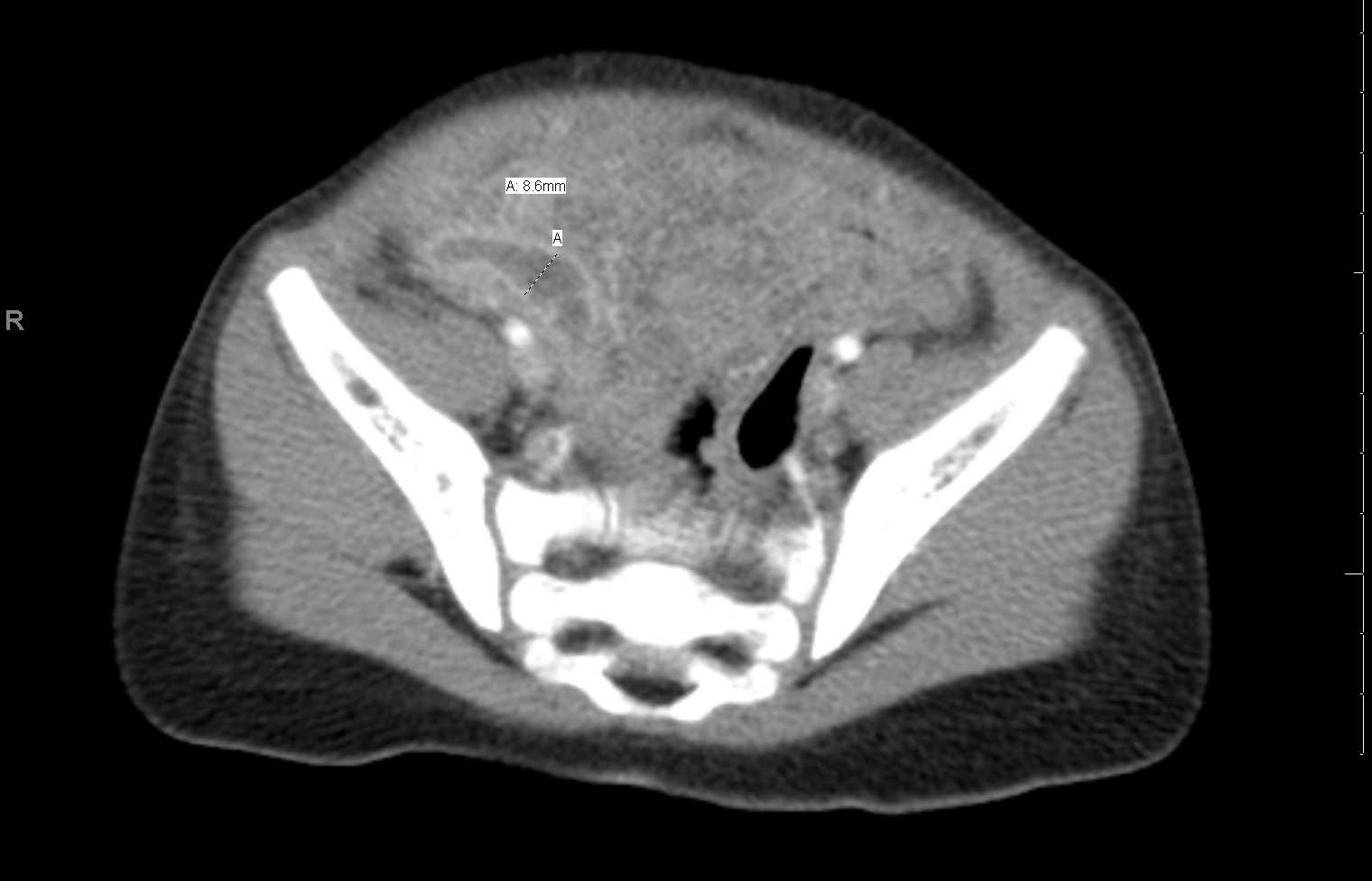
In one meta-analysis, ultrasound has sensitivity and specificity of 69% and 81%, respectively, for the diagnosis of acute appendicitis. 1. CT. CT is highly sensitive (94-98%) and specific (up to 97%) for the diagnosis of acute appendicitis and allows other causes of abdominal pain to be diagnosed. Usually performed with IV contrast (no oral.
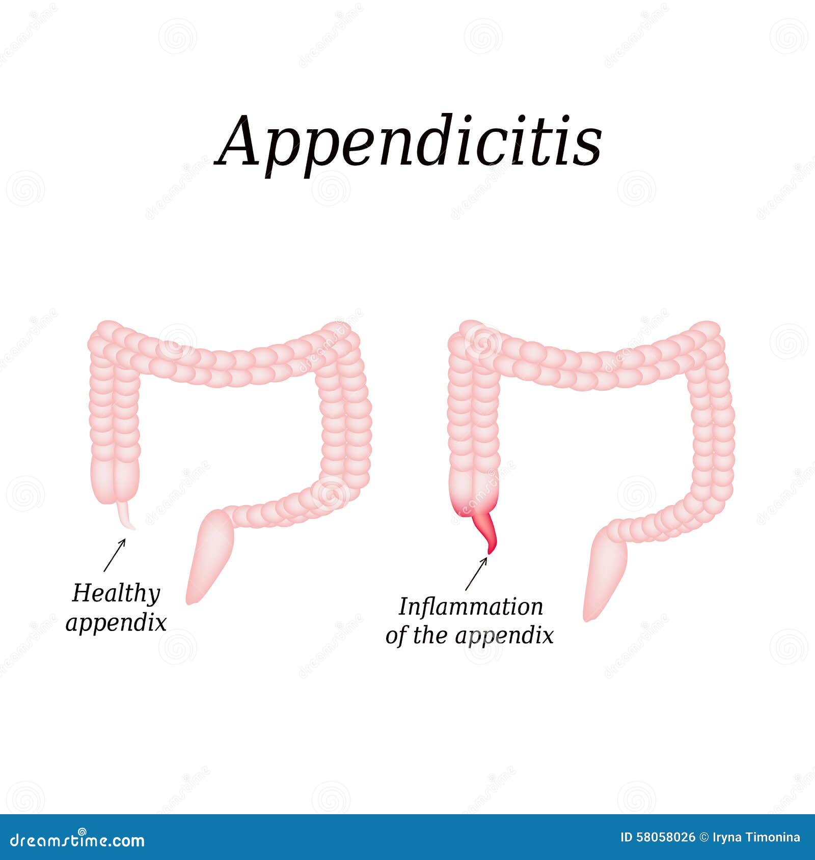
Appendicitis. Inflammation of the Appendix Stock Vector Illustration of care, human 58058026
Penggunaan appendicogram sebagai pemeriksaan penunjang dalam diagnosis appendicitis sudah ditinggalkan, terutama di negara maju. Saat ini telah ada pilihan modalitas pencitraan lain, seperti USG dan CT Scan abdomen, yang lebih mudah dilakukan, memberi hasil lebih cepat, dan lebih tidak invasif. Meski demikian, di Indonesia, pemeriksaan.

Appendicography Stock Image F034/1361 Science Photo Library
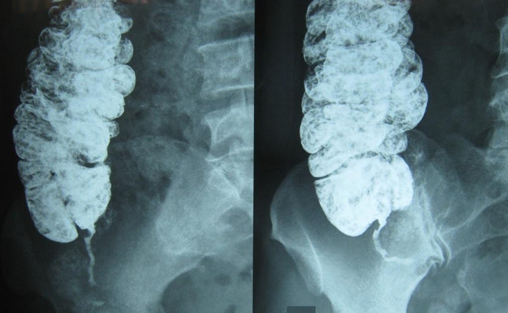
Background: Appendicogram is the barium appendix examination in the form of photo that can help to see blockages or dirt in the appendix. The purpose of this study was to find the Appendicogram.
BLOG HIDUP SEHAT Appendicogram
There are a number of neoplasms that can involve the vermiform appendix, some of which are peculiar to this site.. Epidemiology. Tumors involving the appendix have been found in only about 1% of all appendectomy specimens 9.Epithelial neoplasms and neuroendocrine tumors represent the vast majority of the tumors affecting the appendix.. Clinical presentation

Appendicogram
Reliabilitas Pemeriksaan Appendicogram dalam Penegakan Diagnosis Apendisitis di RSUD Dr. Pirngadi Medan Periode. Jan 2008; M N Hasya; Hasya, M. N. (2012). Reliabilitas Pemeriksaan Appendicogram.
Acute appendicitis sonogram close to the right lower quadrant on... Download Scientific Diagram
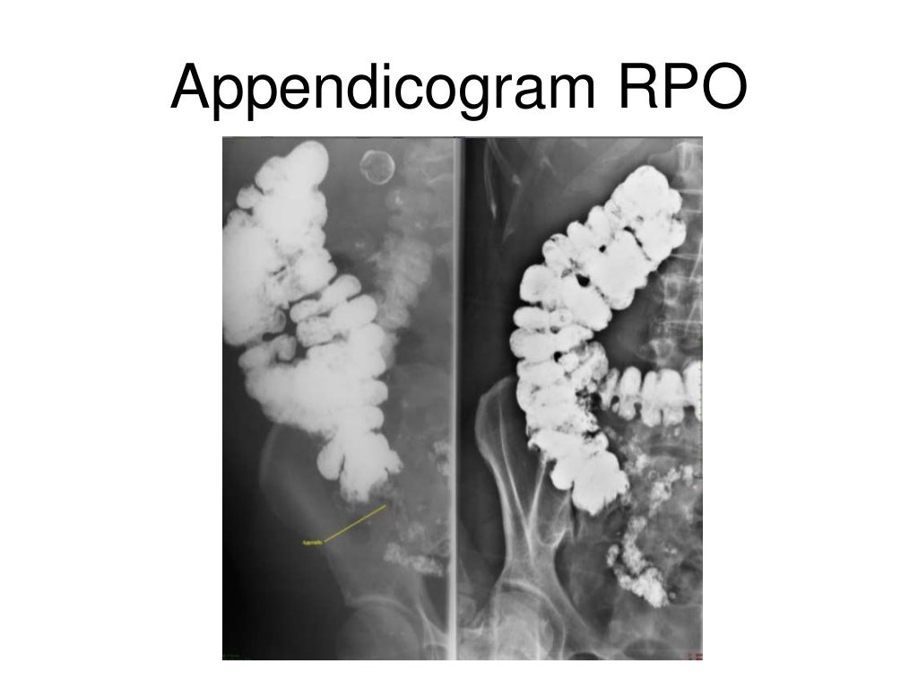
Appendicogram adalah jenis pemeriksaan penunjang radiologi yang digunakan untuk membantu menegakkan diagonosis appendisitis, atau radang usus buntu. Pemeriksaan ini merupakan pemeriksaan yang menggunakan kontras berupa barium sulfat yang diminum sehingga kontras tersebut dapat membantu memvisualisasikan tampakan saluran cerna, mulai dari usus.
Computed tomography showing hyperemic and dilated appendix (red arrow)... Download Scientific
The natural course of untreated appendicitis is reflected in this table. Exact mortality rates in the era before surgery and antibiotics are unknown, but were probably around 10 - 20 %. Nowadays, mortality due to appendicitis has decreased to around 0,1 % , mainly due to early surgery, antibiotics and better diagnosis: US, CT, MRI and also the important lab value CRP.
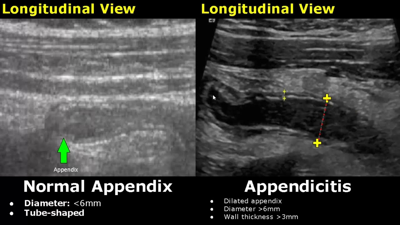
Appendix Ultrasound Normal Vs Abnormal Image Appearances Appendicitis USG Scan YouTube
mengetahui sebab pemilihan appendicogram. Objek dalam penelitian ini yaitu data pasien yang telah menjalani pemeriksaan Appendicogram dengan klinis Apendisitis dengan jumlah pasien sebanyak 2 orang. Alat dan bahan yang digunakan pada penelitian ini adalah lembar daftar wawancara, alat tulis, perekam suara, dan kamera.
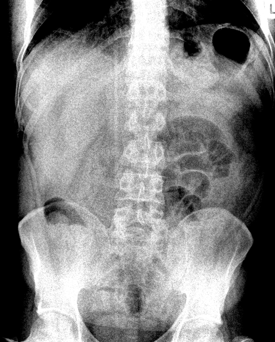
AppendicitisPlain Film Sumer's Radiology Blog
Hasil appendicogram dapat berupa filling (atau disebut juga positive appendicogram), partial filling, ataupun non-filling (negative appendicogram). Hasil pemeriksaan non-filling, artinya cairan kontras tersebut tidak bisa masuk sama sekali ke dalam apendiks atau usus buntu anda. Penyebab dari cairan kontras tersebut tidak bisa masuk adalah.

Classification of acute appendicitis (CAA) type 2b on CT Gangrenous... Download Scientific
Publicationdate 2005-08-14. Introduction. In this overview we focus on nonsurgical appendicitis-mimicking diseases. A correct imaging diagnosis prevents an unnecessary operation or costful in-hospital observation. For critical comments and additional remarks: [email protected]. the Appendix. Normal Appendix.

Carcinoma of the appendix. Axial computed tomography image through the... Download Scientific
Appendicogram dengan klinis Apendisitis di Instalasi Radiologi RSUD Arifin Achmad Provinsi Riau, mengetahui mengapa terjadi perbedaan teknik pemeriksaan appendicogram antara teori dengan RSUD Arifin Achmad Provinsi Riau dan melihat hasil radiograf Appendicogram dengan waktu tunggu 8 jam.

Hasil rontgen appendicogram ("•˘˘•) sick sad ront… Flickr
The appendix is, on average, 8 to 10 cm in length and 7 mm in diameter, with the wall thickness typically 2 mm. [13] Diameter and wall thickness are essential indicators of inflammation where a diameter over 7 mm (over 6 mm in some locations) and a wall thickness greater than 2 to 3 mm are criteria for acute appendicitis when imaging in most.

The Abdominal XRay A relic or a reliable tool? — Taming the SRU
Gross anatomy. The appendix arises from the posteromedial surface of the cecum, approximately 2-3 cm inferior to the ileocecal valve, where the three longitudinal bands of the taeniae coli converge. It is a blind diverticulum which is highly variable in length, ranging between 2 and 20 cm. The appendiceal mesentery is called the mesoappendix 1,2.

Typical Acute Appendicitis Sonography YouTube
Appendicogram is the barium appendix examination in the form of photo that can help to see blockages or dirt in the appendix. The purpose of this study was to find the Appendicogram examination management with clinical appendicitis in the Radiology Installation of Arifin Achmad Hospital, Riau Province, to find out why there were differences in appendicogram examination techniques between the.
The Appendix Radiology Key
Background: Appendicogram is the barium appendix examination in the form of photo that can help to see blockages or dirt in the appendix. The purpose of this study was to find the Appendicogram examination management with clinical appendicitis in the Radiology Installation of Arifin Achmad Hospital, Riau Province, to find out why there were differences in appendicogram examination techniques.